
Search Count: 19
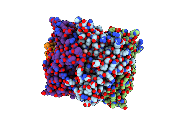 |
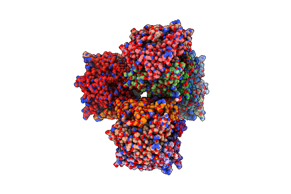
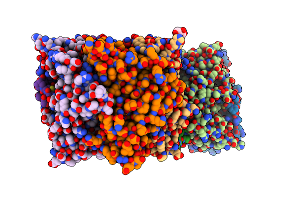
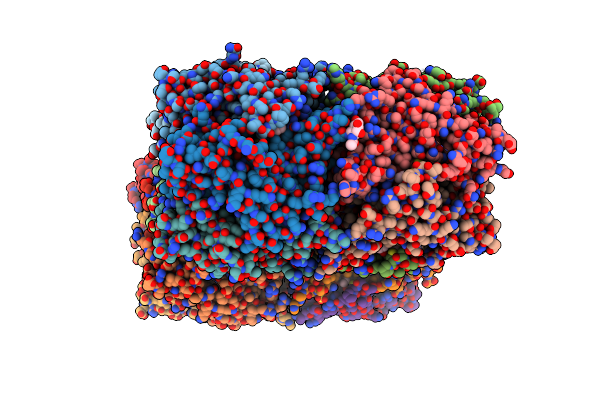
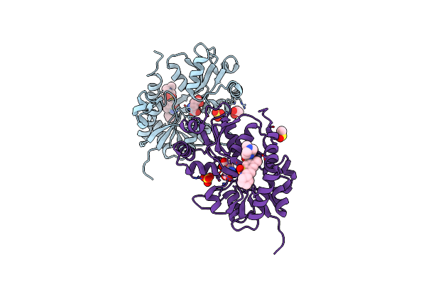
Engineered Ls-Achbp With Alpha4-Alpha4 Binding Pocket In Complex With Ns3573
Organism: Lymnaea stagnalis
Method: X-RAY DIFFRACTION Resolution:2.83 Å Release Date: 2015-07-22 Classification: SIGNALING PROTEIN Ligands: 09P, NAG |
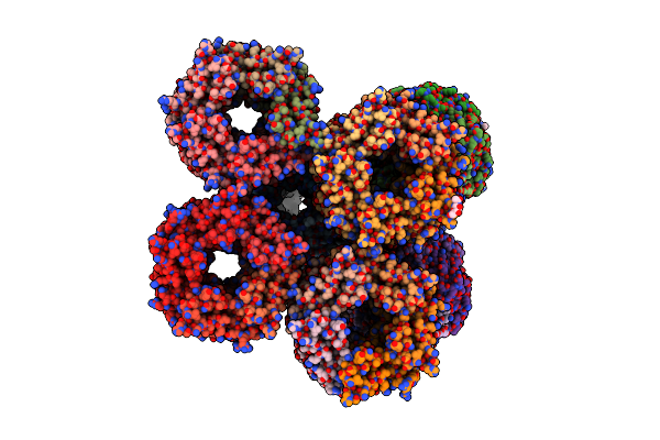 |
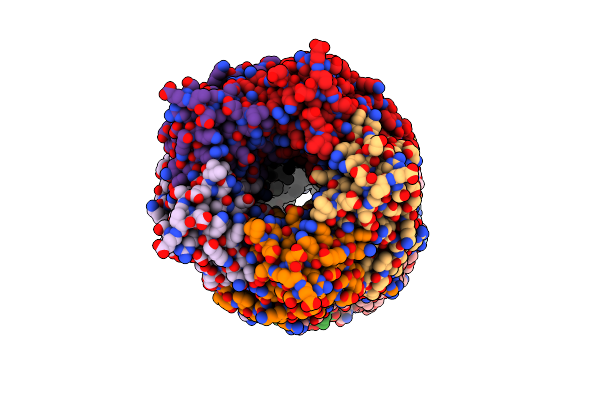
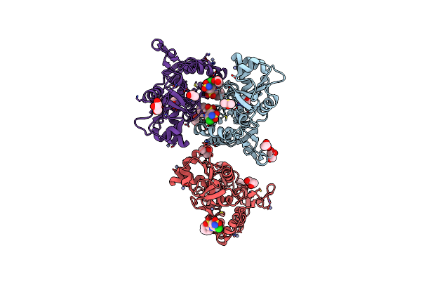
Engineered Ls-Achbp With Alpha4-Alpha4 Binding Pocket In Complex With Ns3920
Organism: Lymnaea stagnalis
Method: X-RAY DIFFRACTION Resolution:2.70 Å Release Date: 2015-07-22 Classification: SIGNALING PROTEIN Ligands: 09R, NAG, SO4 |
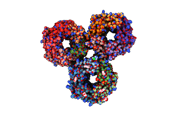 |
Organism: Lymnaea stagnalis
Method: X-RAY DIFFRACTION Resolution:2.68 Å Release Date: 2014-07-09 Classification: ACETYLCHOLINE-BINDING PROTEIN Ligands: 1PE, NAG, SO4, ACT, NSE, CL, GOL |
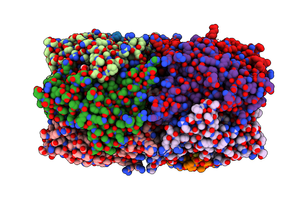 |
Organism: Lymnaea stagnalis
Method: X-RAY DIFFRACTION Resolution:2.60 Å Release Date: 2014-04-23 Classification: ACETYLCHOLINE-BINDING PROTEIN Ligands: ACH, 1PE, SO4, ACT, GOL |
 |
Crystal Structure Of Ls-Achbp Complexed With Carbamoylcholine Analogue 3-(Dimethylamino)Butyl Dimethylcarbamate (Dmabc)
Organism: Lymnaea stagnalis
Method: X-RAY DIFFRACTION Resolution:2.48 Å Release Date: 2013-02-20 Classification: ACETYLCHOLINE-BINDING PROTEIN Ligands: XRX, SO4, NAG, PEG, 1PE |
 |
Crystal Structure Of Ls-Achbp Complexed With Carbamoylcholine Analogue N,N-Dimethyl-4-(1-Methyl-1H-Imidazol-2-Yloxy)Butan-2-Amine
Organism: Lymnaea stagnalis
Method: X-RAY DIFFRACTION Resolution:2.20 Å Release Date: 2013-02-20 Classification: ACETYLCHOLINE-BINDING PROTEIN Ligands: XRS, SO4, NAG |
 |
Crystal Structure Of The Kainate Receptor Gluk3 Ligand Binding Domain In Complex With Kainate
Organism: Rattus norvegicus
Method: X-RAY DIFFRACTION Resolution:2.35 Å Release Date: 2012-05-02 Classification: MEMBRANE PROTEIN Ligands: KAI, CL, K |
 |
Crystal Structure Of The Kainate Receptor Gluk1 Ligand-Binding Domain In Complex With Kainate In The Absence Of Glycerol
Organism: Rattus norvegicus
Method: X-RAY DIFFRACTION Resolution:2.00 Å Release Date: 2012-04-25 Classification: MEMBRANE PROTEIN Ligands: KAI, SO4, CL |
 |
Crystal Structure Of The Acetylcholine Binding Protein (Achbp) From Lymnaea Stagnalis In Complex With Ns3531 (1-(Pyridin-3-Yl)-1,4-Diazepane)
Organism: Lymnaea stagnalis
Method: X-RAY DIFFRACTION Resolution:2.35 Å Release Date: 2011-12-14 Classification: Acetylcholine-binding protein/agonist Ligands: 09O, SO4, NAG |
 |
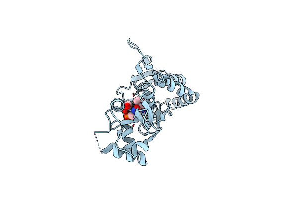
Crystal Structure Of The Acetylcholine Binding Protein (Achbp) From Lymnaea Stagnalis In Complex With Ns3573 (1-(5-Ethoxypyridin-3-Yl)-1,4-Diazepane)
Organism: Lymnaea stagnalis
Method: X-RAY DIFFRACTION Resolution:2.47 Å Release Date: 2011-12-14 Classification: Acetylcholine-binding protein/agonist Ligands: 09P, NAG, SO4 |
 |
Crystal Structure Of The Acetylcholine Binding Protein (Achbp) From Lymnaea Stagnalis In Complex With Ns3570 (1-(5-Phenylpyridin-3-Yl)-1,4-Diazepane)
Organism: Lymnaea stagnalis
Method: X-RAY DIFFRACTION Resolution:2.32 Å Release Date: 2011-12-14 Classification: Acetylcholine-binding protein/agonist Ligands: 09Q, SO4 |
 |
Crystal Structure Of The Acetylcholine Binding Protein (Achbp) From Lymnaea Stagnalis In Complex With Ns3920 (1-(6-Bromopyridin-3-Yl)-1,4-Diazepane)
Organism: Lymnaea stagnalis
Method: X-RAY DIFFRACTION Resolution:2.70 Å Release Date: 2011-12-14 Classification: Acetylcholine-binding protein/agonist Ligands: 09R, SO4 |
 |
Crystal Structure Of The Acetylcholine Binding Protein (Achbp) From Lymnaea Stagnalis In Complex With Ns3950 (1-(6-Bromo-5-Ethoxypyridin-3-Yl)-1,4-Diazepane)
Organism: Lymnaea stagnalis
Method: X-RAY DIFFRACTION Resolution:2.35 Å Release Date: 2011-12-14 Classification: Acetylcholine-binding protein/agonist Ligands: 09S, SO4, NAG |
 |
Crystal Structure Of The Iglur2 Ligand-Binding Core (S1S2J-N754S) In Complex With Glutamate And Cyclothiazide At 2.25 A Resolution
Organism: Rattus norvegicus
Method: X-RAY DIFFRACTION Resolution:2.25 Å Release Date: 2009-07-28 Classification: MEMBRANE PROTEIN Ligands: GLU, CYZ, ZN, GOL, ACT, DMS, CAC |
 |
Crystal Structure Of The Iglur2 Ligand-Binding Core (S1S2J-N754S) In Complex With Glutamate And Ns1493 At 1.85 A Resolution
Organism: Rattus norvegicus
Method: X-RAY DIFFRACTION Resolution:1.85 Å Release Date: 2009-07-28 Classification: MEMBRANE PROTEIN Ligands: GLU, NS3, SO4, GOL, FLC |
 |
Crystal Structure Of The Iglur2 Ligand-Binding Core (S1S2J-N754S) In Complex With Glutamate And Ns5206 At 2.10 A Resolution
Organism: Rattus norvegicus
Method: X-RAY DIFFRACTION Resolution:2.10 Å Release Date: 2009-07-28 Classification: MEMBRANE PROTEIN Ligands: GLU, NS6, GOL, SO4, DMS |
 |
Crystal Structure Of The Iglur2 Ligand-Binding Core (S1S2J-N754S) In Complex With Glutamate And Ns5217 At 1.50 A Resolution
Organism: Rattus norvegicus
Method: X-RAY DIFFRACTION Resolution:1.49 Å Release Date: 2009-07-28 Classification: MEMBRANE PROTEIN Ligands: GLU, NS7, SO4, GOL, DMS |
 |
Crystal Structure Of The Ligand-Binding Core Of Iglur5 In Complex With The Antagonist (S)-Atpo At 1.85 A Resolution
Organism: Rattus norvegicus
Method: X-RAY DIFFRACTION Resolution:1.85 Å Release Date: 2007-07-03 Classification: MEMBRANE PROTEIN Ligands: AT1, GOL |
 |
Crystal Structure Of The Ligand-Binding Core Of Iglur5 In Complex With The Partial Agonist Domoic Acid At 2.5 A Resolution
Organism: Rattus norvegicus
Method: X-RAY DIFFRACTION Resolution:2.50 Å Release Date: 2007-07-03 Classification: MEMBRANE PROTEIN Ligands: DOQ |