
Search Count: 109
 |
Organism: Homo sapiens
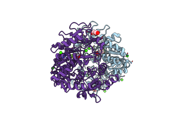
Method: X-RAY DIFFRACTION Release Date: 2025-07-30 Classification: LIGASE Ligands: ZN |
 |
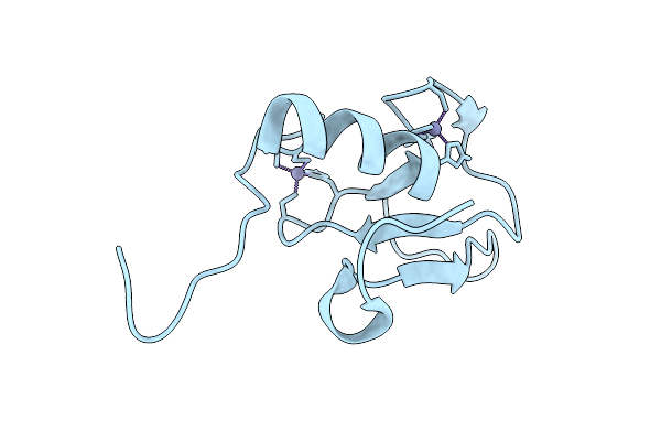
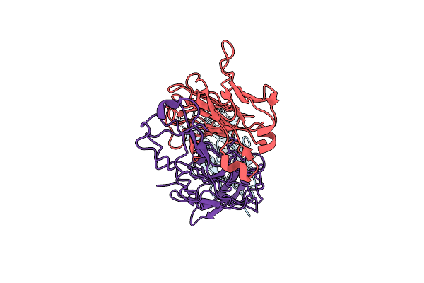
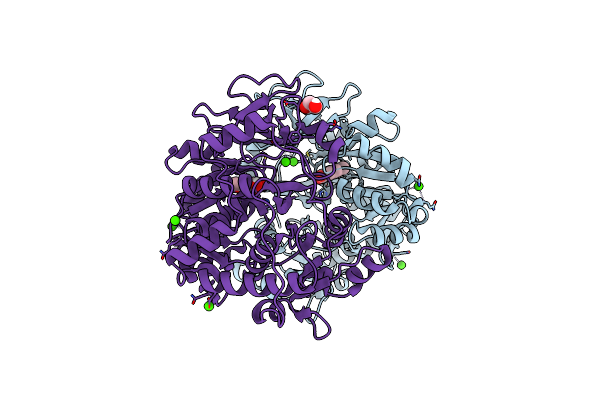
Structure Of Rnf38 Ring With Linchpin Mutant R454Y In Complex With Ubch5B-Ub
Organism: Homo sapiens
Method: X-RAY DIFFRACTION Release Date: 2025-07-30 Classification: LIGASE Ligands: ZN |
 |
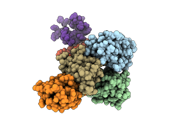
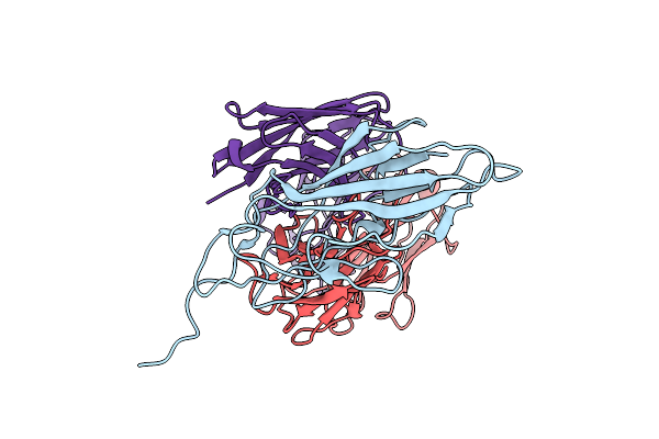
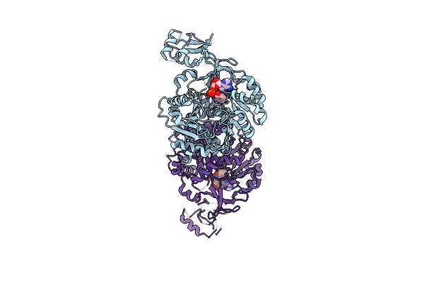
Organism: Escherichia coli uti89, Mus musculus
Method: ELECTRON MICROSCOPY Release Date: 2025-07-02 Classification: CELL ADHESION |
 |
Organism: Escherichia coli uti89, Mus musculus
Method: ELECTRON MICROSCOPY Release Date: 2025-07-02 Classification: CELL ADHESION |
 |
Organism: Escherichia coli uti89, Mus musculus
Method: ELECTRON MICROSCOPY Release Date: 2025-07-02 Classification: CELL ADHESION |
 |
Organism: Escherichia coli uti89, Mus musculus
Method: ELECTRON MICROSCOPY Release Date: 2025-07-02 Classification: CELL ADHESION |
 |
Structure Of The Mouse 8-Oxoguanine Dna Glycosylase Mogg1 In Complex With Ligand Th13579
Organism: Mus musculus
Method: X-RAY DIFFRACTION Release Date: 2025-06-25 Classification: DNA BINDING PROTEIN Ligands: A1ID3, GOL, NI |
 |
Structure Of The Mouse 8-Oxoguanine Dna Glycosylase Mogg1 In Complex With Ligand Th12163
Organism: Mus musculus
Method: X-RAY DIFFRACTION Release Date: 2025-06-25 Classification: DNA BINDING PROTEIN Ligands: A1IEJ, NI |
 |
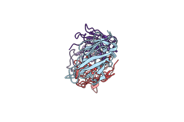
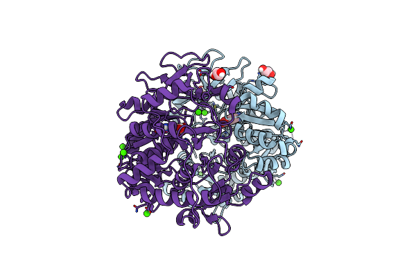
Organism: Homo sapiens
Method: X-RAY DIFFRACTION Release Date: 2025-05-14 Classification: LIGASE Ligands: CA |
 |
Structure Of The Base Mutant V336A, An Nrps Adenylation Domain In The Acinetobactin Biosynthetic Pathway Bound To 4-Methyl Salicylic Acid
Organism: Acinetobacter baumannii
Method: X-RAY DIFFRACTION Resolution:2.39 Å Release Date: 2025-03-26 Classification: LIGASE Ligands: A1BUB, EDO, CA |
 |
Structure Of The Base Double Mutant V336A/S247C, An Nrps Adenylation Domain In The Acinetobactin Biosynthetic Pathway Bound To 4-Fluoro Salicylic Acid
Organism: Acinetobacter baumannii
Method: X-RAY DIFFRACTION Resolution:2.07 Å Release Date: 2025-03-26 Classification: LIGASE Ligands: OOI, PEG, CA, EDO |
 |
Structure Of The Base Mutant V336G, An Nrps Adenylation Domain In The Acinetobactin Biosynthetic Pathway Bound To 4-Amino Salicylic Acid
Organism: Acinetobacter baumannii
Method: X-RAY DIFFRACTION Resolution:2.53 Å Release Date: 2025-03-26 Classification: LIGASE Ligands: BHA, EDO, CA |
 |
Organism: Homo sapiens
Method: X-RAY DIFFRACTION Resolution:1.46 Å Release Date: 2024-12-18 Classification: APOPTOSIS Ligands: A1ILY |
 |
Organism: Homo sapiens
Method: X-RAY DIFFRACTION Resolution:1.42 Å Release Date: 2024-12-18 Classification: APOPTOSIS Ligands: BUD, A1ILV |
 |
Organism: Homo sapiens
Method: X-RAY DIFFRACTION Resolution:1.08 Å Release Date: 2024-12-18 Classification: APOPTOSIS Ligands: A1ILX |
 |
Organism: Homo sapiens
Method: X-RAY DIFFRACTION Resolution:1.39 Å Release Date: 2024-12-18 Classification: APOPTOSIS Ligands: A1ILU |
 |
Organism: Homo sapiens
Method: X-RAY DIFFRACTION Resolution:1.15 Å Release Date: 2024-12-18 Classification: APOPTOSIS Ligands: A1ILW |
 |
Structure Of Fbsh, An Nrps Adenylation Domain In The Fimsbactin Biosynthetic Pathway Bound To 2,3-Dihydroxybenzoic Acid.
Organism: Acinetobacter baumannii
Method: X-RAY DIFFRACTION Resolution:2.30 Å Release Date: 2024-11-20 Classification: LIGASE/SUBSTRATE Ligands: DBH, EDO |
 |
Structure Of Fbsh, An Nrps Adenylation Domain In The Fimsbactin Biosynthetic Pathway Bound To Salicyl-Ams
Organism: Acinetobacter baumannii
Method: X-RAY DIFFRACTION Resolution:2.22 Å Release Date: 2024-11-20 Classification: LIGASE/INHIBITOR Ligands: KT0, EDO |
 |
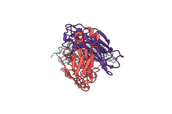
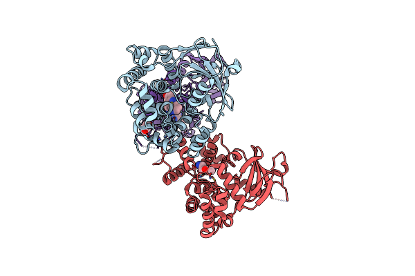
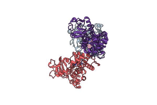
Organism: Homo sapiens
Method: X-RAY DIFFRACTION Release Date: 2024-11-06 Classification: RECOMBINATION Ligands: ADP, BEF, EDO |