
Search Count: 79
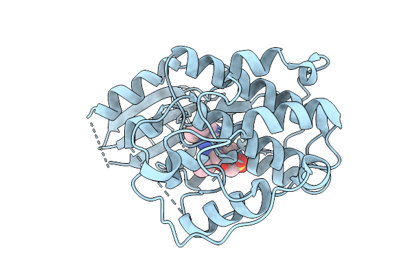 |
Organism: Homo sapiens
Method: X-RAY DIFFRACTION Release Date: 2025-09-10 Classification: TRANSFERASE Ligands: A1INI |
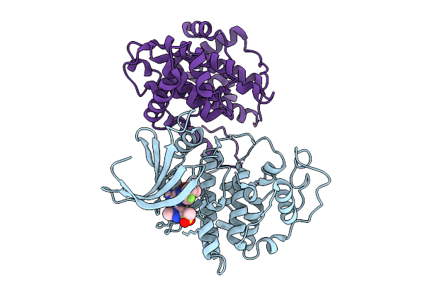 |
Organism: Homo sapiens
Method: X-RAY DIFFRACTION Release Date: 2025-09-10 Classification: TRANSFERASE Ligands: A1IOI |
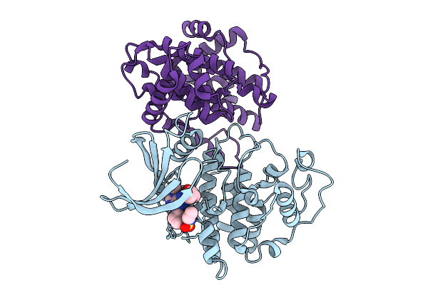 |
Organism: Homo sapiens
Method: X-RAY DIFFRACTION Release Date: 2025-09-10 Classification: TRANSFERASE Ligands: A1IOP |
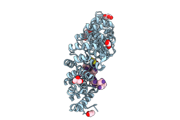 |
Importin Alpha Isoform 2 With Synthetic Zero Net-Charge Nuclear Localization Signal
Organism: Mus musculus, Synthetic construct
Method: X-RAY DIFFRACTION Release Date: 2025-09-03 Classification: PROTEIN TRANSPORT Ligands: GOL, DTT |
 |
Importin Alpha Isoform 2 With Synthetic Zero Net-Charge Nuclear Localization Signal
Organism: Mus musculus, Synthetic construct
Method: X-RAY DIFFRACTION Release Date: 2025-09-03 Classification: PROTEIN TRANSPORT Ligands: GOL, DTT |
 |
Importin Alpha Isoform 2 With Synthetic Zero Net-Charge Nuclear Localization Signal
Organism: Mus musculus, Synthetic construct
Method: X-RAY DIFFRACTION Release Date: 2025-09-03 Classification: PROTEIN TRANSPORT Ligands: GOL, DTT |
 |
Importin Alpha Isoform 2 With Synthetic Zero Net-Charge Nuclear Localization Signal
Organism: Mus musculus, Synthetic construct
Method: X-RAY DIFFRACTION Release Date: 2025-09-03 Classification: PROTEIN TRANSPORT Ligands: GOL, DTT |
 |
Organism: Homo sapiens, Synthetic construct
Method: X-RAY DIFFRACTION Resolution:2.43 Å Release Date: 2024-11-13 Classification: LIPID BINDING PROTEIN Ligands: ZN |
 |
Organism: Homo sapiens
Method: X-RAY DIFFRACTION Resolution:2.43 Å Release Date: 2024-05-29 Classification: TRANSFERASE Ligands: EDO, A1H7X |
 |
Organism: Homo sapiens
Method: X-RAY DIFFRACTION Resolution:1.94 Å Release Date: 2024-05-29 Classification: TRANSFERASE Ligands: EDO, EOH, A1H7Y |
 |
Organism: Homo sapiens
Method: X-RAY DIFFRACTION Resolution:1.70 Å Release Date: 2024-05-29 Classification: TRANSFERASE Ligands: A1H7Z |
 |
Structure Of The Human Ddb1-Dda1-Dcaf15 E3 Ubiquitin Ligase Bound To Compound Furan 12
Organism: Homo sapiens
Method: ELECTRON MICROSCOPY Release Date: 2024-04-03 Classification: LIGASE Ligands: A1H17 |
 |
Structure Of The Human Ddb1-Dda1-Dcaf15 E3 Ubiquitin Ligase Bound To Compound Furan 24
Organism: Homo sapiens
Method: ELECTRON MICROSCOPY Release Date: 2024-04-03 Classification: LIGASE Ligands: A1H18 |
 |
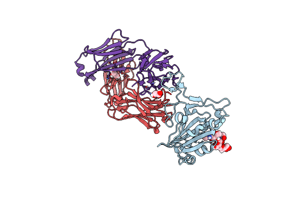
Crystal Structure Of Sars-Cov-2 Receptor Binding Domain In Complex With Neutralizing Human Antibody Wrair-2125.
Organism: Severe acute respiratory syndrome coronavirus 2, Homo sapiens
Method: X-RAY DIFFRACTION Resolution:3.60 Å Release Date: 2021-11-10 Classification: VIRAL PROTEIN/IMMUNE SYSTEM |
 |
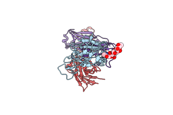
Crystal Structure Of Sars-Cov-2 Receptor Binding Domain In Complex With Neutralizing Human Antibody Wrair-2151.
Organism: Homo sapiens, Severe acute respiratory syndrome coronavirus 2
Method: X-RAY DIFFRACTION Resolution:3.79 Å Release Date: 2021-11-10 Classification: VIRAL PROTEIN/IMMUNE SYSTEM |
 |
Crystal Structure Of Sars-Cov-2 Receptor Binding Domain In Complex With Neutralizing Human Antibody Wrair-2057.
Organism: Homo sapiens, Severe acute respiratory syndrome coronavirus 2
Method: X-RAY DIFFRACTION Resolution:2.28 Å Release Date: 2021-10-06 Classification: VIRAL PROTEIN/IMMUNE SYSTEM Ligands: GOL |
 |
Crystal Structure Of Sars-Cov-2 Receptor Binding Domain In Complex With Neutralizing Human Antibody Wrair-2173.
Organism: Severe acute respiratory syndrome coronavirus 2, Homo sapiens
Method: X-RAY DIFFRACTION Resolution:2.21 Å Release Date: 2021-10-06 Classification: VIRAL PROTEIN/IMMUNE SYSTEM |
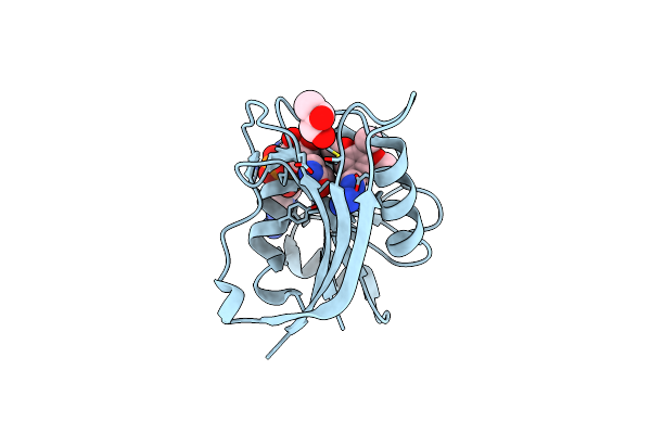 |
X-Ray Structure Of Escherichia Coli Dihydrofolate Reductase L28R Mutant In Complex With Trimethoprim
Organism: Escherichia coli
Method: X-RAY DIFFRACTION Resolution:2.10 Å Release Date: 2021-03-24 Classification: OXIDOREDUCTASE Ligands: NDP, GOL, TOP, CL |
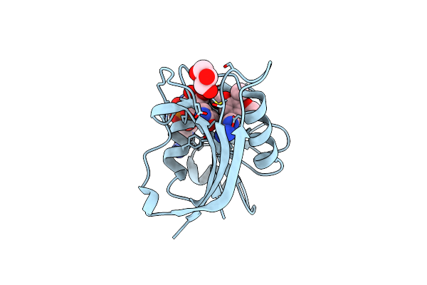 |
X-Ray Structure Of Escherichia Coli Dihydrofolate Reductase In Complex With Trimethoprim
Organism: Escherichia coli
Method: X-RAY DIFFRACTION Resolution:1.90 Å Release Date: 2021-03-24 Classification: OXIDOREDUCTASE Ligands: NDP, GOL, TOP, CL |
 |
Crystal Structure Of Hiv-1 Lm/Hs Clade A/E Crf01 Gp120 Core In Complex With (S)-Mcg-Iv-210
Organism: Human immunodeficiency virus 1
Method: X-RAY DIFFRACTION Resolution:2.50 Å Release Date: 2020-10-07 Classification: IMMUNE SYSTEM Ligands: NAG, O51, EPE |