
Search Count: 5
 |
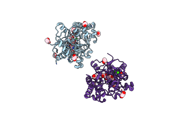
Structure Of The Periplasmic Binding Protein (Pbp) Occj From A. Tumefaciens B6 In Complex With Octopine.
Organism: Agrobacterium tumefaciens str. b6
Method: X-RAY DIFFRACTION Resolution:1.99 Å Release Date: 2017-12-20 Classification: Octopine-binding protein Ligands: EDO, NA, CL, GOL, 6DB, ACT |
 |
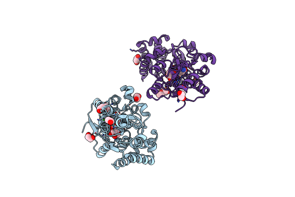
Crystal Structure Of The Pbp Mota In Complex With Mannopine From A. Tumefaciens B6
Organism: Agrobacterium tumefaciens str. b6
Method: X-RAY DIFFRACTION Resolution:1.75 Å Release Date: 2016-09-21 Classification: TRANSPORT PROTEIN Ligands: MO0, EDO, CA |
 |
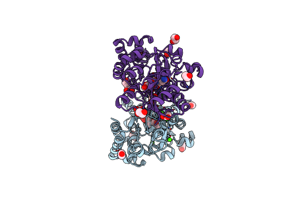
Crystal Structure Of The Periplasmic Binding Protein Mota In Complex With Dfg From A. Tumefaciens B6
Organism: Agrobacterium tumefaciens str. b6
Method: X-RAY DIFFRACTION Resolution:1.90 Å Release Date: 2016-09-21 Classification: TRANSPORT PROTEIN Ligands: SNW, PEG, EDO, CA |
 |
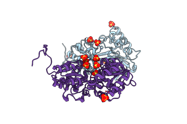
Crystal Structure Of The Pbp Mota From A. Tumefaciens B6 In Complex With Glucopine
Organism: Agrobacterium tumefaciens str. b6
Method: X-RAY DIFFRACTION Resolution:1.80 Å Release Date: 2016-09-21 Classification: TRANSPORT PROTEIN Ligands: GOP, CA, EDO |
 |
Organism: Agrobacterium tumefaciens str. b6
Method: X-RAY DIFFRACTION Resolution:2.54 Å Release Date: 2016-09-21 Classification: TRANSPORT PROTEIN Ligands: SO4 |