
Search Count: 16
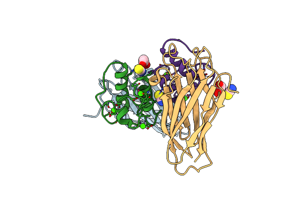 |
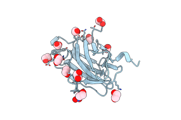
Crystal Structure Of The Sixth Cohesin From Acetivibrio Cellulolyticus' Scaffoldin B In Complex With Cel5 Dockerin S15I, I16N Mutant
Organism: Acetivibrio cellulolyticus
Method: X-RAY DIFFRACTION Resolution:1.45 Å Release Date: 2018-01-31 Classification: PROTEIN BINDING Ligands: CA, SCN, GOL |
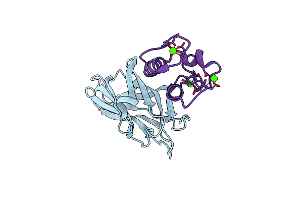 |
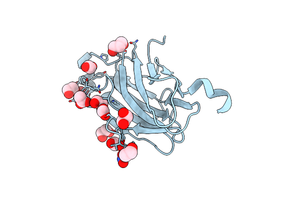
Crystal Structure Of The Sixth Cohesin From Acetivibrio Cellulolyticus' Scaffoldin B In Complex With Cel5 Dockerin S51I, L52N Mutant
Organism: Acetivibrio cellulolyticus
Method: X-RAY DIFFRACTION Resolution:1.40 Å Release Date: 2018-01-31 Classification: CELL ADHESION Ligands: CA |
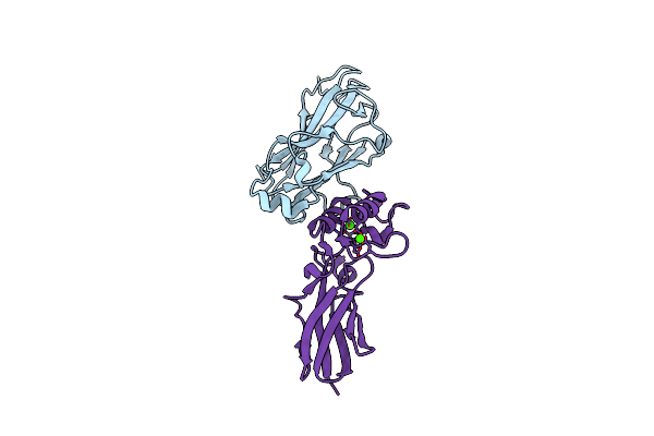 |
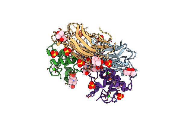
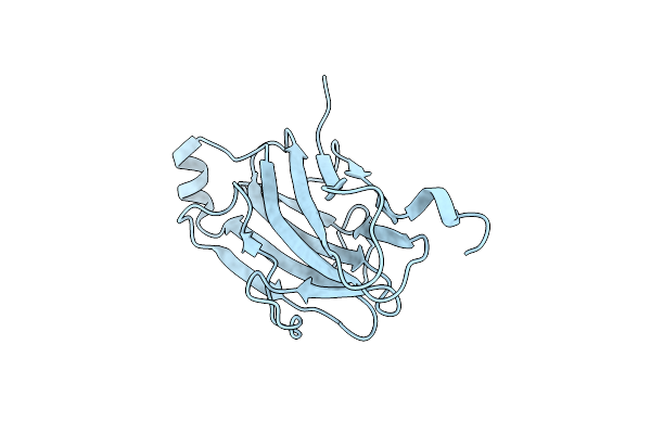
Crystal Structure Of Coh3Scab-Xdoc_M2Scaa Complex: A C-Terminal Interface Mutant Of Type Ii Cohesin-X-Dockerin Complex From Acetivibrio Cellulolyticus
Organism: Acetivibrio cellulolyticus
Method: X-RAY DIFFRACTION Resolution:1.93 Å Release Date: 2015-10-07 Classification: STRUCTURAL PROTEIN Ligands: CA |
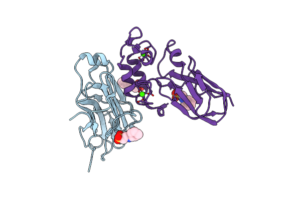 |
Crystal Structure Of Coh3Scab-Xdoc_M1Scaa Complex: A N-Terminal Interface Mutant Of Type Ii Cohesin-X-Dockerin Complex From Acetivibrio Cellulolyticus
Organism: Acetivibrio cellulolyticus
Method: X-RAY DIFFRACTION Resolution:1.64 Å Release Date: 2015-07-29 Classification: PROTEIN BINDING Ligands: NHE, CA |
 |
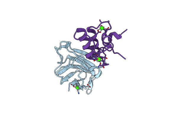
High Resolution Structure Of The Third Cohesin Scac In Complex With The Scab Dockerin With A Mutation In The N-Terminal Helix (In To Si) From Acetivibrio Cellulolyticus Displaying A Type I Interaction.
Organism: Acetivibrio cellulolyticus
Method: X-RAY DIFFRACTION Resolution:1.49 Å Release Date: 2015-04-15 Classification: CELL ADHESION/PROTEIN BINDING Ligands: SO4, EPE, MPD, CA |
 |
High Resolution Structure Of The Third Cohesin Scac In Complex With The Scab Dockerin With A Mutation In The C-Terminal Helix (In To Si) From Acetivibrio Cellulolyticus Displaying A Type I Interaction.
Organism: Acetivibrio cellulolyticus
Method: X-RAY DIFFRACTION Resolution:1.81 Å Release Date: 2015-04-15 Classification: CELL ADHESION/PROTEIN BINDING Ligands: CA |
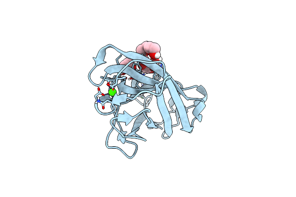 |
Structure Of Cbm3B Of Major Scaffoldin Subunit Scaa From Acetivibrio Cellulolyticus
Organism: Acetivibrio cellulolyticus
Method: X-RAY DIFFRACTION Resolution:1.07 Å Release Date: 2012-01-11 Classification: CARBOHYDRATE-BINDING PROTEIN Ligands: CA, EDO, NI, 1PE |
 |
Structure Of Cbm3B Of Major Scaffoldin Subunit Scaa From Acetivibrio Cellulolyticus Determined On The Nikel Absorption Edge
Organism: Acetivibrio cellulolyticus
Method: X-RAY DIFFRACTION Resolution:1.80 Å Release Date: 2012-01-11 Classification: SUGAR BINDING PROTEIN Ligands: CA, EDO, NI, 1PE |
 |
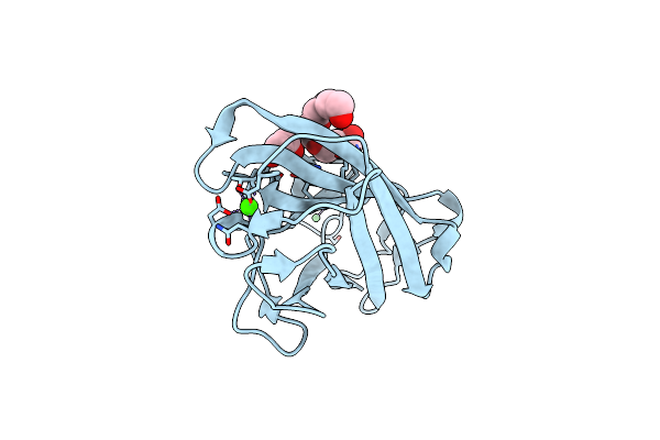
Structure Of Cbm3B Of Major Scaffoldin Subunit Scaa From Acetivibrio Cellulolyticus Determined From The Crystals Grown In The Presence Of Nickel
Organism: Acetivibrio cellulolyticus
Method: X-RAY DIFFRACTION Resolution:1.00 Å Release Date: 2012-01-11 Classification: CRYSTALLINE CELLULOSE-BINDING PROTEIN Ligands: CA, EDO, NI, 1PE |
 |
Structure Analysis Of The Type Ii Cohesin Dyad From The Adaptor Scaa Scaffoldin Of Acetivibrio Cellulolyticus
Organism: Acetivibrio cellulolyticus
Method: X-RAY DIFFRACTION Resolution:1.57 Å Release Date: 2010-05-05 Classification: STRUCTURAL PROTEIN, PROTEIN BINDING Ligands: EDO, PDO, HEZ |
 |
Crystal Structure Of The Second Type Ii Cohesin Module From The Cellulosomal Adaptor Scaa Scaffoldin Of Acetivibrio Cellulolyticus
Organism: Acetivibrio cellulolyticus
Method: X-RAY DIFFRACTION Resolution:1.99 Å Release Date: 2009-06-23 Classification: STRUCTURAL PROTEIN Ligands: EDO, ACT, PDO, BU1 |
 |
Structure Of The Second Type Ii Cohesin Module From The Adaptor Scaa Scaffoldin Of Acetivibrio Cellulolyticus (Including Long C-Terminal Linker)
Organism: Acetivibrio cellulolyticus
Method: X-RAY DIFFRACTION Resolution:2.49 Å Release Date: 2009-06-23 Classification: STRUCTURAL PROTEIN Ligands: EDO |
 |
Crystal Structure Of The Type Ii Cohesin Module From The Cellulosome Of Acetivibrio Cellulolyticus With An Extended Linker Conformation
Organism: Acetivibrio cellulolyticus
Method: X-RAY DIFFRACTION Resolution:1.20 Å Release Date: 2009-01-13 Classification: STRUCTURAL PROTEIN Ligands: NO3, CL, EDO |
 |
Crystal Structure Analysis Of The K171A Mutation Of N-Terminal Type Ii Cohesin 1 From The Cellulosomal Scab Subunit Of Acetivibrio Cellulolyticus
Organism: Acetivibrio cellulolyticus
Method: X-RAY DIFFRACTION Resolution:1.85 Å Release Date: 2008-12-02 Classification: STRUCTURAL PROTEIN Ligands: EDO, NH4, NO3 |
 |
Crystal Structure Analysis Of A Type Ii Cohesin Domain From The Cellulosome Of Acetivibrio Cellulolyticus- Semet Derivative
Organism: Acetivibrio cellulolyticus
Method: X-RAY DIFFRACTION Resolution:1.28 Å Release Date: 2006-06-13 Classification: cellulosome Ligands: NO3, EDO, PDO, FMT, ACY |
 |
Crystal Structure Analysis Of A Type Ii Cohesin Domain From The Cellulosome Of Acetivibrio Cellulolyticus
Organism: Acetivibrio cellulolyticus
Method: X-RAY DIFFRACTION Resolution:1.90 Å Release Date: 2004-09-21 Classification: STRUCTURAL PROTEIN |