
Search Count: 64
 |
Organism: Acanthamoeba polyphaga mimivirus
Method: X-RAY DIFFRACTION Resolution:2.10 Å Release Date: 2025-08-06 Classification: VIRAL PROTEIN Ligands: UDP, MN |
 |
Organism: Acanthamoeba polyphaga mimivirus
Method: X-RAY DIFFRACTION Resolution:1.80 Å Release Date: 2025-06-11 Classification: VIRAL PROTEIN Ligands: MN |
 |
Organism: Acanthamoeba polyphaga mimivirus
Method: X-RAY DIFFRACTION Resolution:1.75 Å Release Date: 2025-06-11 Classification: VIRAL PROTEIN Ligands: UDP, MN, GOL |
 |
Organism: Acanthamoeba polyphaga mimivirus
Method: X-RAY DIFFRACTION Resolution:1.50 Å Release Date: 2025-06-11 Classification: VIRAL PROTEIN Ligands: UDP, MN, BGC, GAL |
 |
Organism: Acanthamoeba polyphaga mimivirus
Method: X-RAY DIFFRACTION Resolution:2.30 Å Release Date: 2025-06-04 Classification: VIRAL PROTEIN |
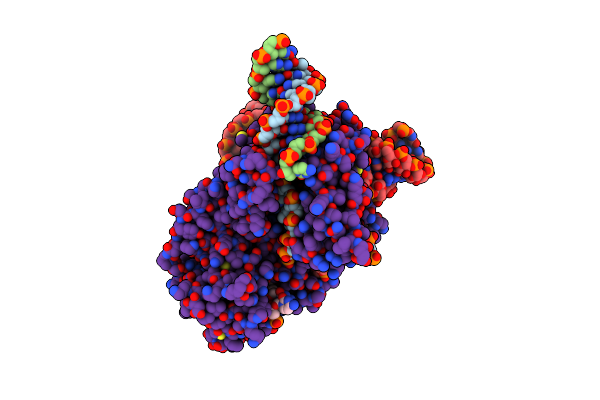 |
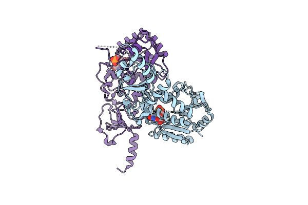
Cryo-Em Structure Of Acanthamoeba Polyphaga Mimivirus Fanzor2 Ternary Complex
Organism: Acanthamoeba polyphaga mimivirus
Method: ELECTRON MICROSCOPY Release Date: 2024-10-09 Classification: DNA BINDING PROTEIN/DNA/RNA Ligands: ZN, MG |
 |
Organism: Acanthamoeba polyphaga mimivirus
Method: ELECTRON MICROSCOPY Release Date: 2022-08-10 Classification: VIRAL PROTEIN Ligands: FAD |
 |
Organism: Acanthamoeba polyphaga mimivirus
Method: ELECTRON MICROSCOPY Release Date: 2022-08-10 Classification: VIRAL PROTEIN Ligands: FAD |
 |
Organism: Acanthamoeba polyphaga mimivirus
Method: ELECTRON MICROSCOPY Release Date: 2022-08-10 Classification: VIRAL PROTEIN Ligands: FAD |
 |
Organism: Acanthamoeba polyphaga mimivirus
Method: ELECTRON MICROSCOPY Release Date: 2022-08-10 Classification: VIRAL PROTEIN Ligands: FAD |
 |
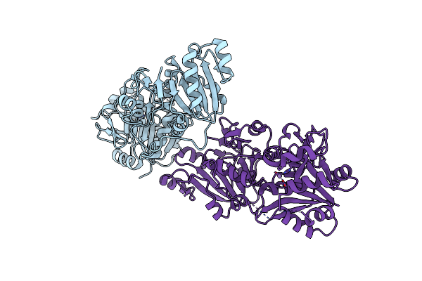
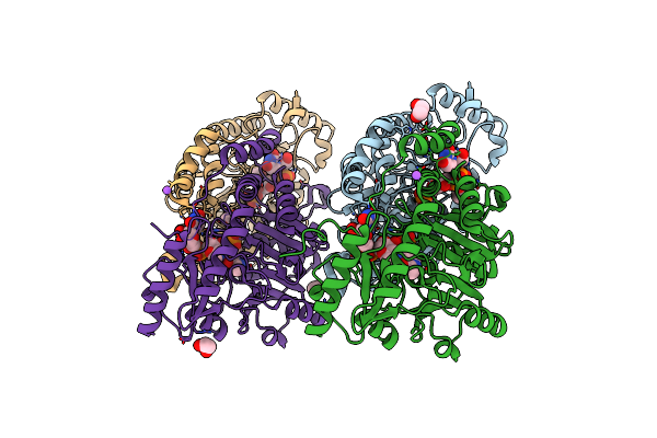
X-Ray Structure Of The L136 Aminotransferase From Acanthamoeba Polyphaga Mimivirus In The Presence Of Tdp And Pmp
Organism: Acanthamoeba polyphaga mimivirus
Method: X-RAY DIFFRACTION Resolution:1.70 Å Release Date: 2021-04-28 Classification: TRANSFERASE Ligands: PMP, PO4, EDO, CL, TYD |
 |
Crystal Structure Of The L136 Aminotransferase K185A From Acanthamoeba Polyphaga Mimivirus In The Presence Of The Udp-Viosamine External Aldimine
Organism: Acanthamoeba polyphaga mimivirus
Method: X-RAY DIFFRACTION Resolution:1.85 Å Release Date: 2021-04-28 Classification: TRANSFERASE Ligands: Z7P, CL, EDO, NA |
 |
Crystal Structure Of The L136 Aminotransferase From Acanthamoeba Polyphaga Mimivirus In Complex With The Tdp-Viosamine External Aldimine
Organism: Acanthamoeba polyphaga mimivirus
Method: X-RAY DIFFRACTION Resolution:1.95 Å Release Date: 2021-04-28 Classification: TRANSFERASE Ligands: T4K, CL, EDO, NA |
 |
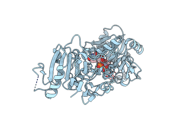
Crystal Structure Of The L780 Udp-Rhamnose Synthase From Acanthamoeba Polyphaga Mimivirus
Organism: Acanthamoeba polyphaga mimivirus
Method: X-RAY DIFFRACTION Resolution:1.45 Å Release Date: 2020-08-26 Classification: OXIDOREDUCTASE Ligands: AWU, NAP, EDO |
 |
Organism: Acanthamoeba polyphaga mimivirus, Bacillus phage pbs2
Method: X-RAY DIFFRACTION Resolution:2.60 Å Release Date: 2020-07-08 Classification: DNA BINDING PROTEIN/INHIBITOR |
 |
Organism: Acanthamoeba polyphaga mimivirus, Bacillus phage pbs2
Method: X-RAY DIFFRACTION Resolution:3.10 Å Release Date: 2020-07-08 Classification: DNA BINDING PROTEIN/INHIBITOR |
 |
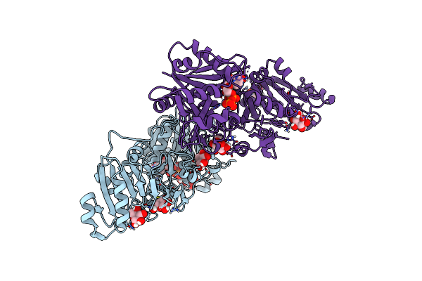
X-Ray Structure Of The R141 Sugar 4,6-Dehydratase From Acanthamoeba Polyphaga Minivirus
Organism: Acanthamoeba polyphaga mimivirus
Method: X-RAY DIFFRACTION Resolution:2.05 Å Release Date: 2020-03-04 Classification: OXIDOREDUCTASE Ligands: TYD, NAD, NI |
 |
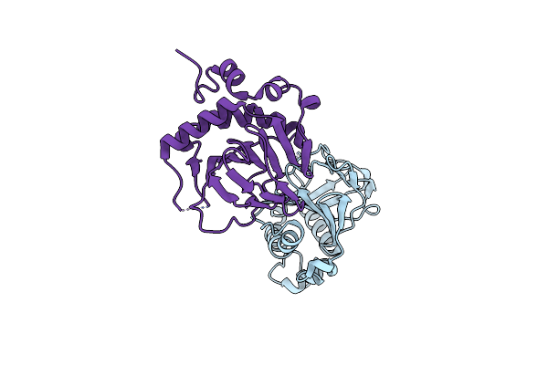
Structural And Mechanistic Analyses Reveal A Unique Cas4-Like Protein In The Mimivirus Virophage Resistance Element System
Organism: Acanthamoeba polyphaga mimivirus
Method: X-RAY DIFFRACTION Resolution:3.00 Å Release Date: 2018-07-25 Classification: NUCLEAR PROTEIN Ligands: MG |
 |
Organism: Acanthamoeba polyphaga mimivirus
Method: X-RAY DIFFRACTION Resolution:2.81 Å Release Date: 2018-06-20 Classification: NUCLEAR PROTEIN |
 |
The Crystal Structure Of A Lysyl Hydroxylase From Acanthamoeba Polyphaga Mimivirus
Organism: Acanthamoeba polyphaga mimivirus
Method: X-RAY DIFFRACTION Resolution:2.24 Å Release Date: 2018-02-21 Classification: OXIDOREDUCTASE Ligands: IOD, FE2 |