
Search Count: 5
 |
Organism: Corynebacterium diphtheriae
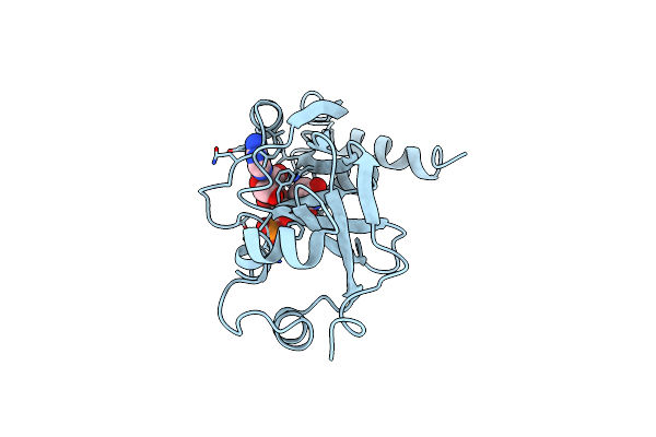
Method: X-RAY DIFFRACTION Resolution:2.10 Å Release Date: 2023-07-05 Classification: TOXIN Ligands: APU |
 |
Organism: Corynebacterium diphtheriae
Method: X-RAY DIFFRACTION Resolution:1.55 Å Release Date: 2000-11-16 Classification: TOXIN Ligands: CL, APU |
 |
Organism: Corynephage beta
Method: X-RAY DIFFRACTION Resolution:2.50 Å Release Date: 1994-11-01 Classification: TOXIN Ligands: APU |
 |
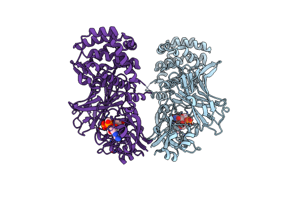
The Refined Structure Of Dimeric Diphtheria Toxin At 2.0 Angstroms Resolution
Organism: Corynephage beta
Method: X-RAY DIFFRACTION Resolution:2.00 Å Release Date: 1994-07-31 Classification: TOXIN Ligands: APU |
 |
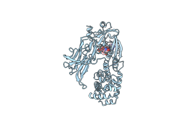
The Refined Structure Of Monomeric Diphtheria Toxin At 2.3 Angstroms Resolution
Organism: Corynephage beta
Method: X-RAY DIFFRACTION Resolution:2.30 Å Release Date: 1994-07-31 Classification: TOXIN Ligands: APU |