
Search Count: 830
 |
Organism: Escherichia coli
Method: ELECTRON MICROSCOPY Release Date: 2026-01-07 Classification: DNA BINDING PROTEIN/RNA/DNA Ligands: ATP, MG |
 |
Organism: Escherichia coli
Method: ELECTRON MICROSCOPY Release Date: 2025-12-31 Classification: DNA BINDING PROTEIN/RNA/DNA Ligands: ATP, MG |
 |
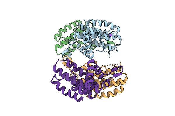
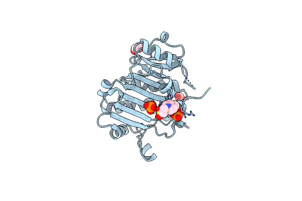
Mycobacterium Tuberculosis Relbe1 Toxin-Antitoxin System; Rv1247C (Relb1 Antitoxin), Rv1246C (Rele1 Toxin)
Organism: Mycobacterium tuberculosis h37rv
Method: X-RAY DIFFRACTION Release Date: 2025-12-17 Classification: TOXIN Ligands: CL |
 |
Organism: Escherichia coli
Method: ELECTRON MICROSCOPY Release Date: 2025-12-03 Classification: DNA BINDING PROTEIN Ligands: ZN |
 |
Organism: Escherichia coli
Method: ELECTRON MICROSCOPY Release Date: 2025-11-19 Classification: ANTITOXIN/DNA/RNA |
 |
Organism: Escherichia coli
Method: ELECTRON MICROSCOPY Release Date: 2025-11-19 Classification: ANTITOXIN/DNA/RNA Ligands: ATP |
 |
Organism: Escherichia coli k-12, Synthetic construct
Method: X-RAY DIFFRACTION Release Date: 2025-10-29 Classification: TOXIN |
 |
Organism: Bacteroides fragilis
Method: X-RAY DIFFRACTION Release Date: 2025-10-01 Classification: ANTITOXIN Ligands: IPA |
 |
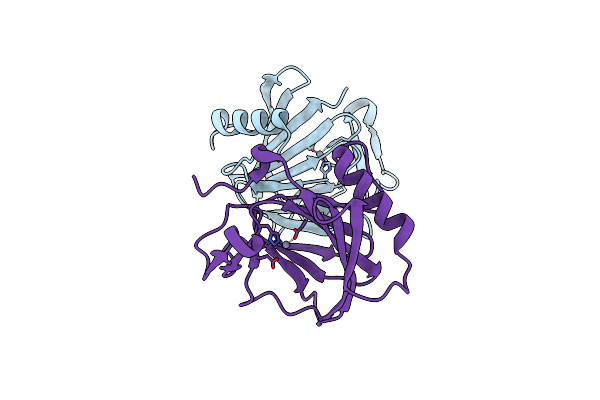
Crystal Structure Of Neutralizing Fab Eq4.Dp46-3D From Equine Antivenom Bound To A Consensus Short Chain Three Finger Alpha-Neurotoxin.
Organism: Synthetic construct, Equus caballus
Method: X-RAY DIFFRACTION Release Date: 2025-09-10 Classification: ANTITOXIN |
 |
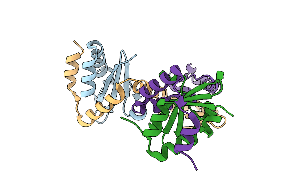
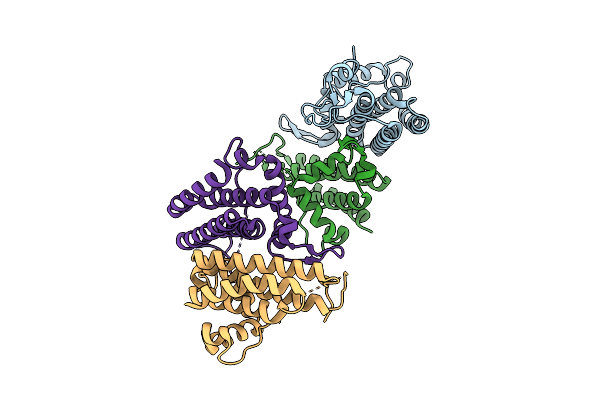
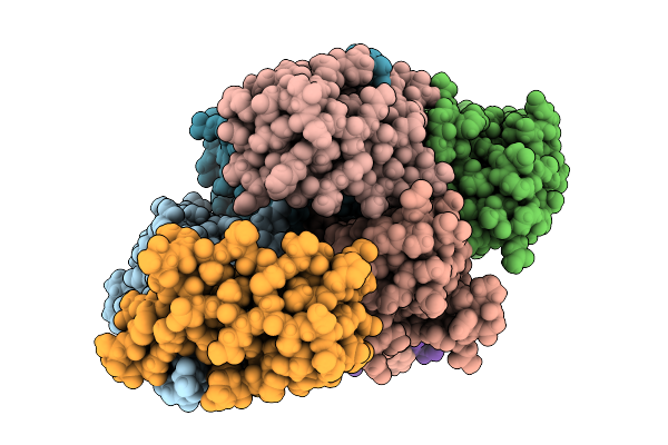
Crystal Structure Of The Pseudomonas Putida Xre-Res Toxin-Antitoxin Complex Bound To Promoter Dna
Organism: Pseudomonas putida kt2440
Method: X-RAY DIFFRACTION Release Date: 2025-08-20 Classification: GENE REGULATION |
 |
Organism: Lactococcus cremoris
Method: X-RAY DIFFRACTION Release Date: 2025-08-13 Classification: RNA BINDING PROTEIN |
 |
Crystal Structure Of The Hepn Family Member Abiv, An Rnase In A Two-Component Antiphage System In Lactococcus Lactis
Organism: Lactococcus cremoris
Method: X-RAY DIFFRACTION Release Date: 2025-08-13 Classification: RNA BINDING PROTEIN |
 |
Organism: Lactococcus cremoris
Method: X-RAY DIFFRACTION Release Date: 2025-08-13 Classification: RNA BINDING PROTEIN Ligands: SO4 |
 |
Organism: Vicugna pacos, Naja kaouthia
Method: X-RAY DIFFRACTION Release Date: 2025-08-06 Classification: TOXIN Ligands: HOH |
 |
Organism: Vibrio cholerae serotype o1 (strain atcc 39315 / el tor inaba n16961)
Method: X-RAY DIFFRACTION Release Date: 2025-07-30 Classification: TOXIN Ligands: SO4, ACT |
 |
Organism: Proteus vulgaris
Method: X-RAY DIFFRACTION Release Date: 2025-07-02 Classification: ANTITOXIN/DNA BINDING PROTEIN/DNA Ligands: MG, CL |
 |
Organism: Proteus vulgaris
Method: X-RAY DIFFRACTION Release Date: 2025-07-02 Classification: ANTITOXIN/DNA BINDING PROTEIN/DNA Ligands: MG |
 |
Organism: Salmonella enterica subsp. enterica serovar typhimurium
Method: X-RAY DIFFRACTION Release Date: 2025-07-02 Classification: STRUCTURAL PROTEIN Ligands: ZN |
 |
Crystal Structure Of The Immunity Protein (52 Domain-Containing Protein) From Pseudomonas Aeruginosa Pao1.
Organism: Pseudomonas aeruginosa pao1
Method: X-RAY DIFFRACTION Release Date: 2025-06-18 Classification: ANTITOXIN Ligands: EPE, PO4, EDO, CO3 |
 |
Organism: Mycobacterium tuberculosis (strain cdc 1551 / oshkosh)
Method: X-RAY DIFFRACTION Release Date: 2025-06-11 Classification: TOXIN |