
Search Count: 8
 |
Organism: Synthetic construct
Method: X-RAY DIFFRACTION Release Date: 2025-10-15 Classification: DE NOVO PROTEIN Ligands: ALL |
 |
Organism: Synthetic construct
Method: X-RAY DIFFRACTION Release Date: 2025-10-15 Classification: DE NOVO PROTEIN Ligands: ALL |
 |
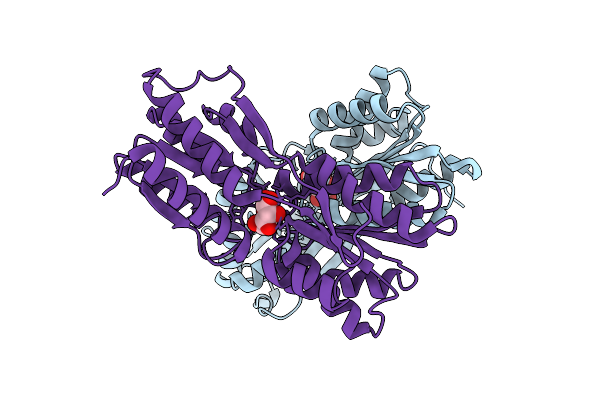
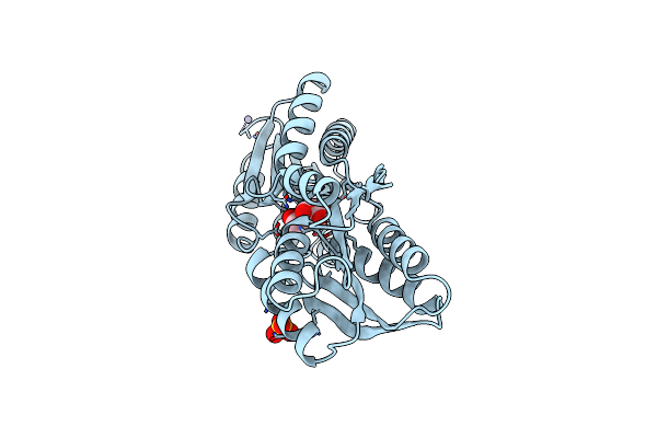
Crystal Structure Of Lactobacillus Rhamnosus L-Rhamnose Isomerase In Complex With D-Allose
Organism: Lacticaseibacillus rhamnosus
Method: X-RAY DIFFRACTION Resolution:1.71 Å Release Date: 2024-03-13 Classification: ISOMERASE Ligands: MN, ALL, AFD |
 |
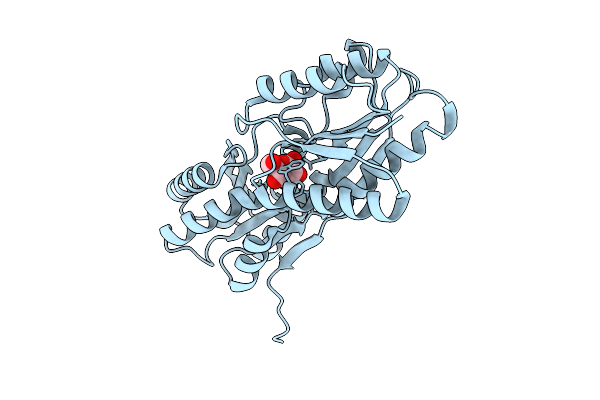
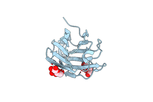
X-Ray Structure Of Enterobacter Cloacae Allose-Binding Protein In Complex With D-Allose
Organism: Enterobacter cloacae
Method: X-RAY DIFFRACTION Resolution:1.74 Å Release Date: 2023-10-25 Classification: SUGAR BINDING PROTEIN Ligands: ALL |
 |
Organism: Homo sapiens
Method: X-RAY DIFFRACTION Resolution:1.92 Å Release Date: 2020-03-04 Classification: SUGAR BINDING PROTEIN Ligands: ALL |
 |
Organism: Homo sapiens
Method: SOLUTION NMR Release Date: 2016-06-08 Classification: ISOMERASE Ligands: ALL |
 |
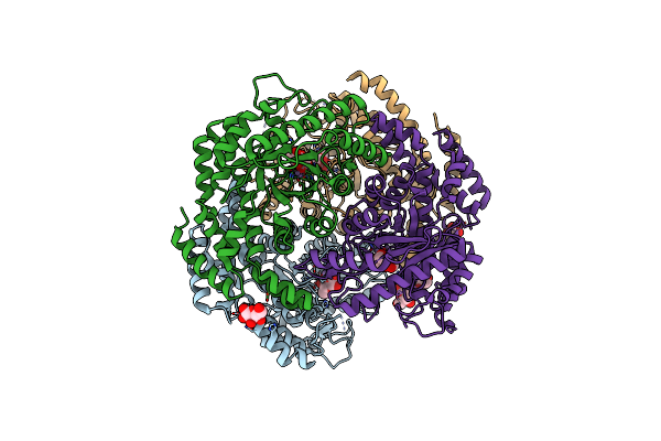
Crystal Structure Of An Abc Transporter Periplasmic Solute Binding Protein (Ipr025997) From Actinobacillus Succinogenes 130Z(Asuc_0081, Target Efi-511065) With Bound D-Allose
Organism: Actinobacillus succinogenes (strain atcc 55618 / 130z)
Method: X-RAY DIFFRACTION Resolution:2.70 Å Release Date: 2015-10-07 Classification: TRANSPORT PROTEIN Ligands: ALL |
 |
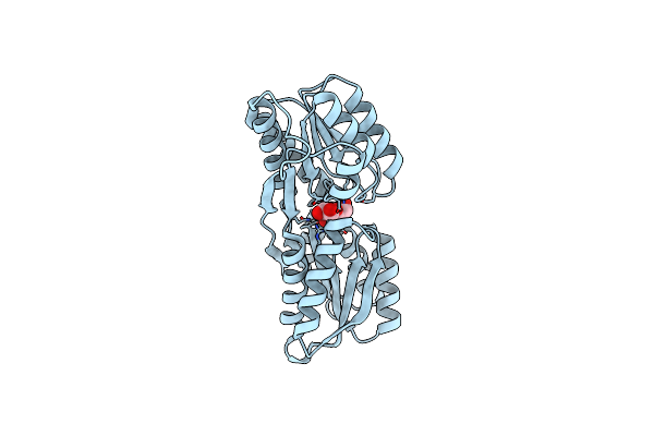
Organism: Escherichia coli k12
Method: X-RAY DIFFRACTION Resolution:1.80 Å Release Date: 1999-02-16 Classification: PERIPLASMIC SUGAR RECEPTOR Ligands: ZN, SO4, ALL |