Search Count: 3,450
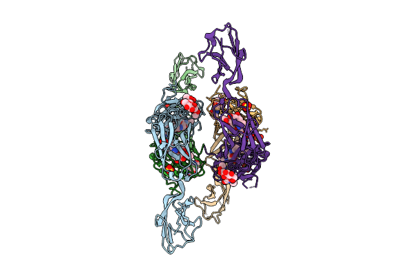 |
Crystal Structure Of Human Cd22 Ig Domains 1-3 In Complex With Modified Sialoside 7-012
Organism: Homo sapiens
Method: X-RAY DIFFRACTION Release Date: 2025-12-10 Classification: IMMUNE SYSTEM Ligands: A1JJO, SO4 |
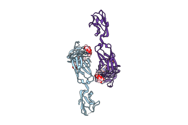 |
Crystal Structure Of Human Cd22 Ig Domains 1-3 In Complex With Modified Sialoside 1B
Organism: Homo sapiens
Method: X-RAY DIFFRACTION Release Date: 2025-12-10 Classification: IMMUNE SYSTEM Ligands: A1JJN, GOL |
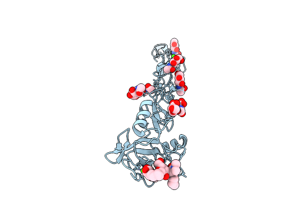 |
Organism: Homo sapiens
Method: X-RAY DIFFRACTION Release Date: 2025-12-03 Classification: CELL ADHESION Ligands: NAG, CA, A1IUQ |
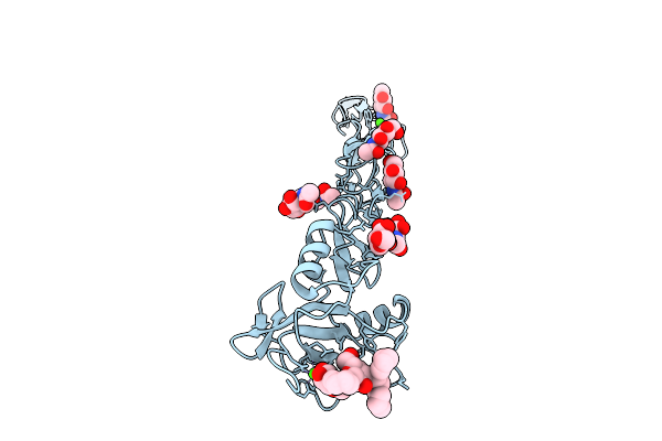 |
Organism: Homo sapiens
Method: X-RAY DIFFRACTION Release Date: 2025-12-03 Classification: CELL ADHESION Ligands: NAG, A1IUR, CA |
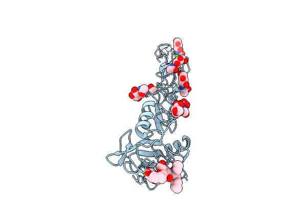 |
Organism: Homo sapiens
Method: X-RAY DIFFRACTION Release Date: 2025-12-03 Classification: CELL ADHESION Ligands: NAG, A1IUS, CA |
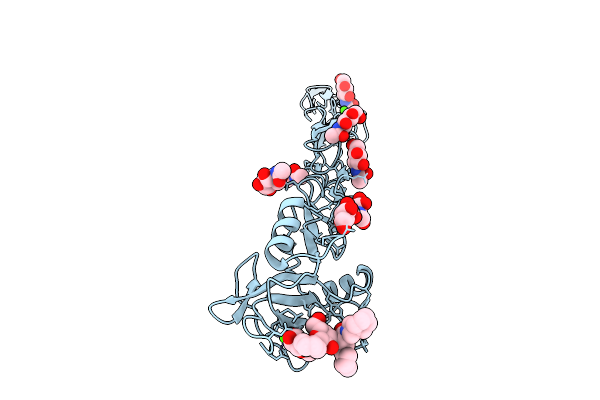 |
Organism: Homo sapiens
Method: X-RAY DIFFRACTION Release Date: 2025-12-03 Classification: CELL ADHESION Ligands: NAG, A1IUT, CA |
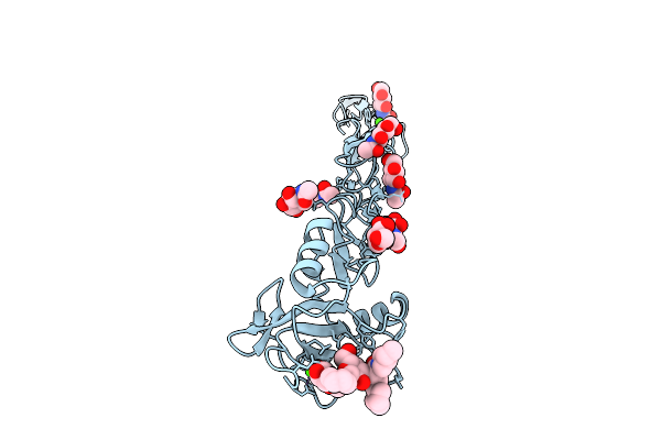 |
Organism: Homo sapiens
Method: X-RAY DIFFRACTION Release Date: 2025-12-03 Classification: CELL ADHESION Ligands: NAG, A1IUU, CA |
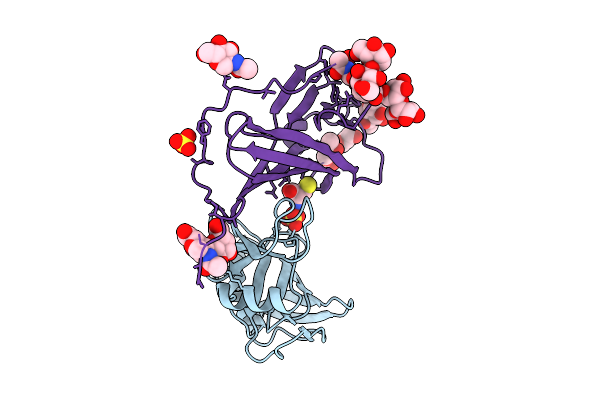 |
Organism: Mus musculus, Murid betaherpesvirus 1
Method: X-RAY DIFFRACTION Release Date: 2025-11-26 Classification: VIRAL PROTEIN Ligands: SO4, NAG, CYS |
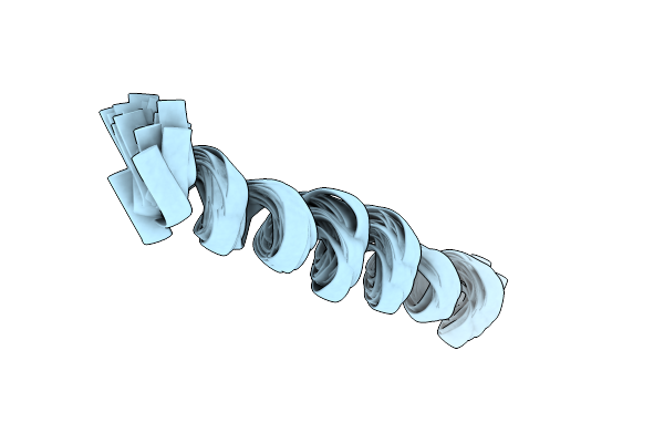 |
Solution Nmr Structure Of A Peptide Encompassing Residues 967-991 Of The Human Formin Inf2
|
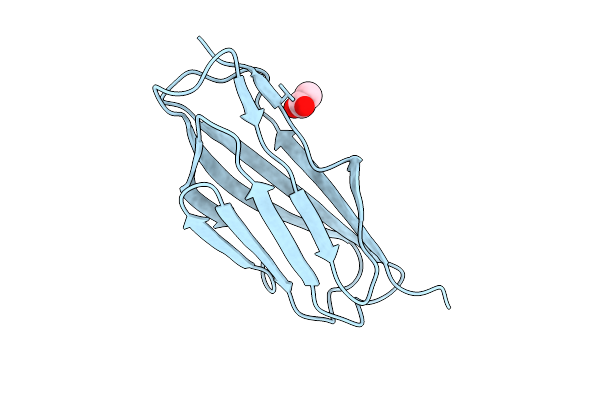 |
Organism: Acinetobacter silvestris
Method: X-RAY DIFFRACTION Release Date: 2025-11-26 Classification: CELL ADHESION Ligands: ACT |
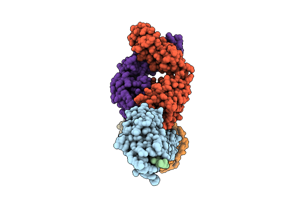 |
Antibody Fragments From Mab475 And Mab824 Bound To The Adhesin Protein Fimh
Organism: Escherichia coli, Escherichia coli k-12, Mus musculus
Method: ELECTRON MICROSCOPY Release Date: 2025-11-26 Classification: CELL ADHESION/IMMUNE SYSTEM |
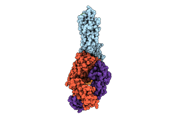 |
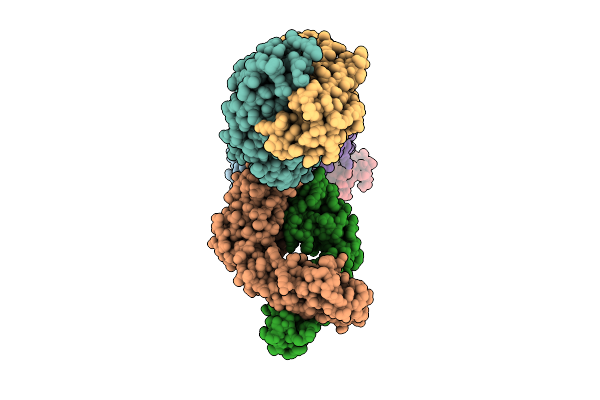
Antibody Fragments From Mab824 And Mab926 Bound To The Adhesin Protein Fimh
Organism: Escherichia coli, Mus musculus
Method: ELECTRON MICROSCOPY Release Date: 2025-11-26 Classification: CELL ADHESION/IMMUNE SYSTEM |
 |
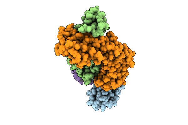
Antibody Fragments From Mab21, Mab475, And Mab824 Bound To The Adhesin Protein Fimh
Organism: Escherichia coli, Mus musculus
Method: ELECTRON MICROSCOPY Release Date: 2025-11-26 Classification: CELL ADHESION/IMMUNE SYSTEM |
 |
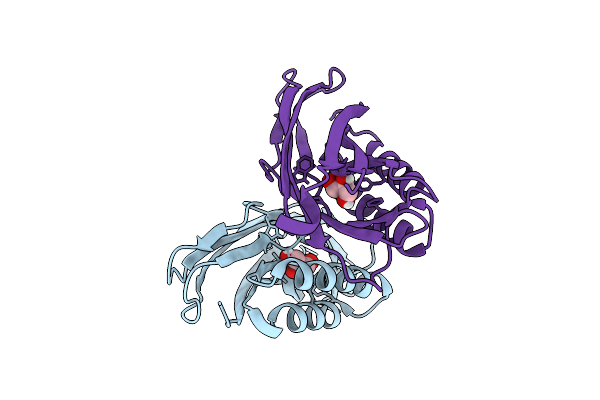
Antibody Fragments From Mab21 And Mab824 Bound To The Adhesin Protein Fimh Containing Alpha-Methyl Mannose
Organism: Escherichia coli, Mus musculus
Method: ELECTRON MICROSCOPY Release Date: 2025-11-26 Classification: CELL ADHESION/IMMUNE SYSTEM Ligands: MMA |
 |
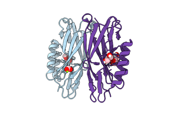
Organism: Escherichia coli
Method: X-RAY DIFFRACTION Release Date: 2025-11-26 Classification: CELL ADHESION Ligands: GOL |
 |
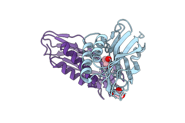
Organism: Escherichia coli
Method: X-RAY DIFFRACTION Release Date: 2025-11-26 Classification: CELL ADHESION Ligands: GOL, FMT, MG |
 |
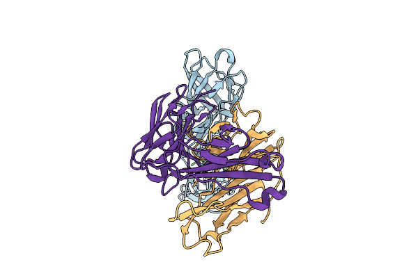
Organism: Escherichia coli
Method: X-RAY DIFFRACTION Release Date: 2025-11-26 Classification: CELL ADHESION Ligands: GOL |
 |
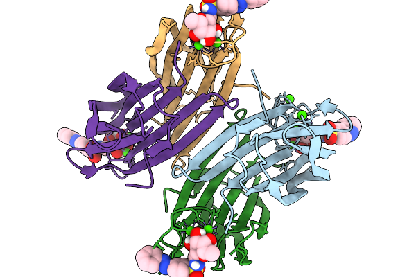
Organism: Escherichia coli, Mus musculus
Method: ELECTRON MICROSCOPY Release Date: 2025-11-26 Classification: CELL ADHESION/IMMUNE SYSTEM |
 |
Organism: Pseudomonas aeruginosa
Method: X-RAY DIFFRACTION Release Date: 2025-11-19 Classification: SUGAR BINDING PROTEIN Ligands: A1IT5, CA |
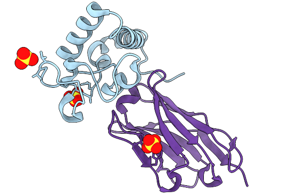 |
Crystal Structure Of Tetraspanin Cd63Mutant Large Extracellular Loop (Lel) In Complex With Sybody Sb3
Organism: Homo sapiens, Synthetic construct
Method: X-RAY DIFFRACTION Release Date: 2025-11-19 Classification: CELL ADHESION Ligands: SO4 |

