
Search Count: 704
All
Selected
 |
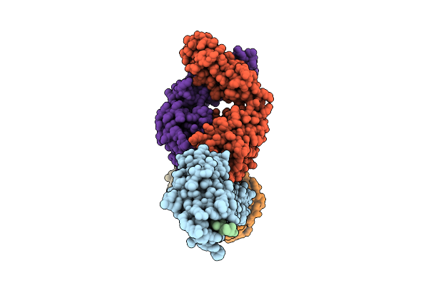
Bordetella Filamentous Hemagglutinin (Fhab) C-Terminal Domain Bound To Microtubules
Organism: Bordetella bronchiseptica rb50, Sus scrofa
Method: ELECTRON MICROSCOPY Release Date: 2026-02-25 Classification: TOXIN Ligands: GDP, TA1, GTP, MG |
 |
Organism: Aequorea victoria, Escherichia phage pp01
Method: X-RAY DIFFRACTION Resolution:2.10 Å Release Date: 2026-01-07 Classification: CELL ADHESION Ligands: CA |
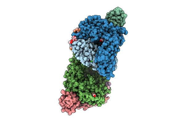 |
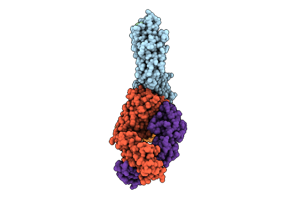
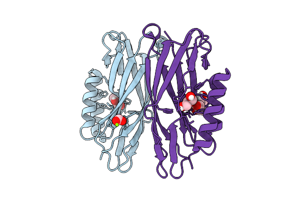
Vibrio Cholerae Protein Frha Peptid-Binding Domain And Adjacent Split Domain (S1127-F1439) In Complex With Peptide Agwtd X-Ray Crystallography Structure
Organism: Vibrio cholerae o395, Vibrio cholerae
Method: X-RAY DIFFRACTION Resolution:2.40 Å Release Date: 2025-12-03 Classification: METAL BINDING PROTEIN Ligands: CA, PEG, EDO |
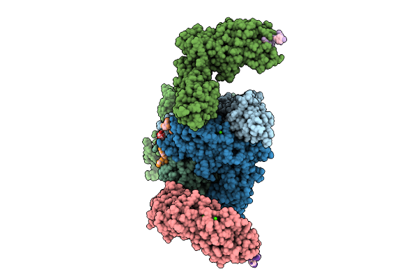 |
Vibrio Cholerae Protein Frha Peptid-Binding Domain And Adjacent Split Domain (S1127-F1439) In Complex With Peptide Agytd X-Ray Crystallography Structure
Organism: Vibrio cholerae o395, Vibrio cholerae
Method: X-RAY DIFFRACTION Resolution:2.50 Å Release Date: 2025-12-03 Classification: METAL BINDING PROTEIN Ligands: CA |
 |
Organism: Acinetobacter silvestris
Method: X-RAY DIFFRACTION Resolution:1.77 Å Release Date: 2025-11-26 Classification: CELL ADHESION Ligands: ACT |
 |
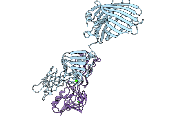
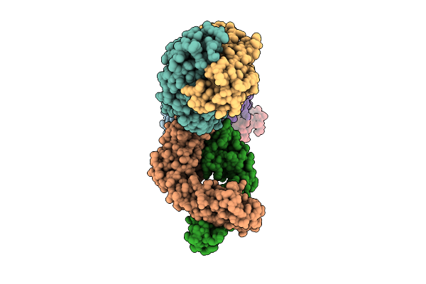
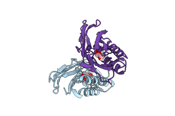
Antibody Fragments From Mab475 And Mab824 Bound To The Adhesin Protein Fimh
Organism: Escherichia coli, Escherichia coli k-12, Mus musculus
Method: ELECTRON MICROSCOPY Resolution:3.20 Å Release Date: 2025-11-26 Classification: CELL ADHESION/IMMUNE SYSTEM |
 |
Antibody Fragments From Mab824 And Mab926 Bound To The Adhesin Protein Fimh
Organism: Escherichia coli, Mus musculus
Method: ELECTRON MICROSCOPY Release Date: 2025-11-26 Classification: CELL ADHESION/IMMUNE SYSTEM |
 |
Antibody Fragments From Mab21, Mab475, And Mab824 Bound To The Adhesin Protein Fimh
Organism: Escherichia coli, Mus musculus
Method: ELECTRON MICROSCOPY Resolution:3.20 Å Release Date: 2025-11-26 Classification: CELL ADHESION/IMMUNE SYSTEM |
 |
Antibody Fragments From Mab21 And Mab824 Bound To The Adhesin Protein Fimh Containing Alpha-Methyl Mannose
Organism: Escherichia coli, Mus musculus
Method: ELECTRON MICROSCOPY Release Date: 2025-11-26 Classification: CELL ADHESION/IMMUNE SYSTEM Ligands: MMA |
 |
Organism: Escherichia coli
Method: X-RAY DIFFRACTION Resolution:1.70 Å Release Date: 2025-11-26 Classification: CELL ADHESION Ligands: GOL |
 |
Organism: Escherichia coli
Method: X-RAY DIFFRACTION Resolution:1.65 Å Release Date: 2025-11-26 Classification: CELL ADHESION Ligands: GOL, FMT, MG |
 |
Organism: Escherichia coli
Method: X-RAY DIFFRACTION Resolution:1.68 Å Release Date: 2025-11-26 Classification: CELL ADHESION Ligands: GOL |
 |
Organism: Escherichia coli, Mus musculus
Method: ELECTRON MICROSCOPY Release Date: 2025-11-26 Classification: CELL ADHESION/IMMUNE SYSTEM |
 |
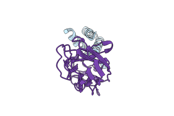
Organism: Streptococcus
Method: X-RAY DIFFRACTION Resolution:1.29 Å Release Date: 2025-11-26 Classification: SUGAR BINDING PROTEIN Ligands: CA, NA |
 |
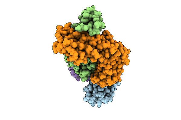
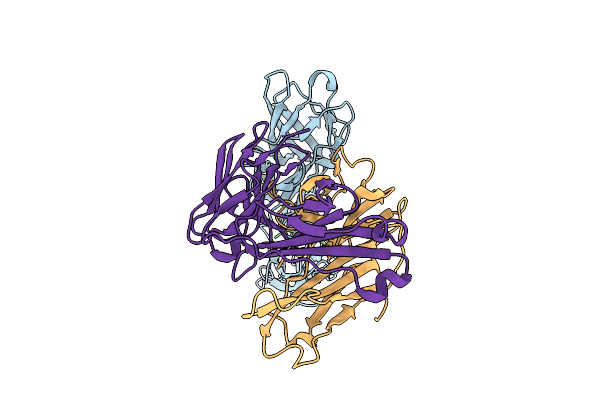
Crystal Structure Of The Transmembrane Domain Of Trimeric Autotransporter Adhesin Ataa In Complex With The N-Terminal Domain Of Tpga
Organism: Acinetobacter sp. tol 5
Method: X-RAY DIFFRACTION Resolution:2.60 Å Release Date: 2025-10-08 Classification: CELL ADHESION Ligands: CA, C8E, PGE, MES, CL |
 |
Crystal Structure Of Aeromonas Salmonicida Putative Carbohydrate Binding Module And Split Domain
Organism: Aeromonas salmonicida subsp. salmonicida a449
Method: X-RAY DIFFRACTION Resolution:2.50 Å Release Date: 2025-09-24 Classification: CELL ADHESION Ligands: CA |
 |
In Situ Cryo-Em Structure Of Outer Membrane Cap (Omc) Of The Legionella Dot/Icm T4Ss Machine
Organism: Legionella pneumophila subsp. pneumophila
Method: ELECTRON MICROSCOPY Release Date: 2025-09-17 Classification: PROTEIN TRANSPORT |
 |
Organism: Synthetic construct, Acinetobacter baumannii acicu
Method: X-RAY DIFFRACTION Resolution:1.35 Å Release Date: 2025-09-17 Classification: PROTEIN BINDING |
 |
Organism: Synthetic construct, Escherichia coli
Method: X-RAY DIFFRACTION Resolution:1.75 Å Release Date: 2025-09-17 Classification: DE NOVO PROTEIN |
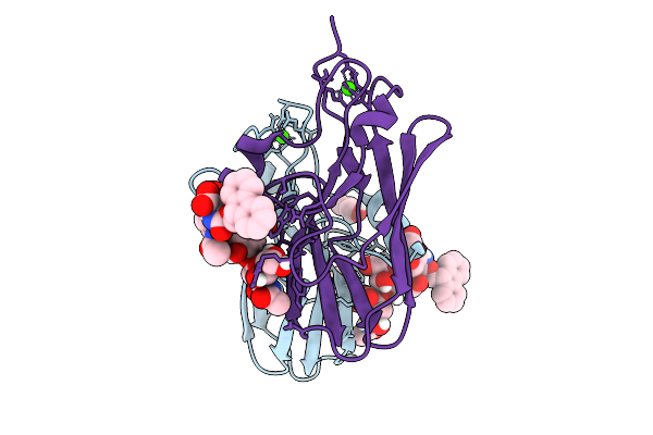 |
Organism: Streptococcus
Method: X-RAY DIFFRACTION Resolution:1.89 Å Release Date: 2025-09-10 Classification: SUGAR BINDING PROTEIN Ligands: GOL, VP1, SER, CA |