
Search Count: 35
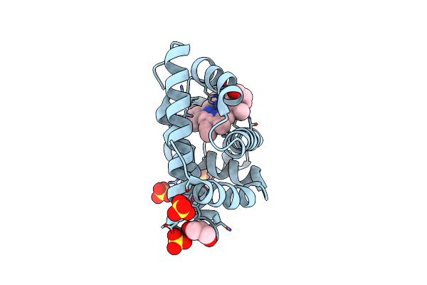 |
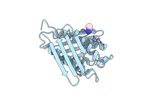
Organism: Physeter catodon
Method: X-RAY DIFFRACTION Resolution:1.76 Å Release Date: 2023-06-21 Classification: OXYGEN STORAGE Ligands: HEM, ACM, GOL, SO4 |
 |
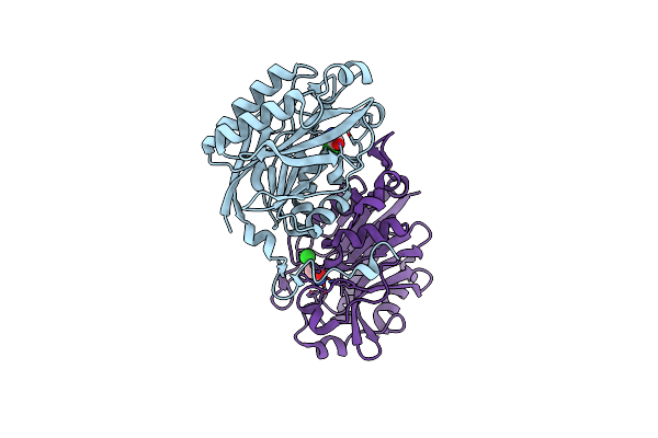
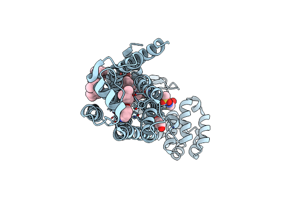
Organism: Homo sapiens, Synthetic construct
Method: X-RAY DIFFRACTION Resolution:2.69 Å Release Date: 2022-09-14 Classification: TRANSCRIPTION Ligands: V7J, D5M, ACM |
 |
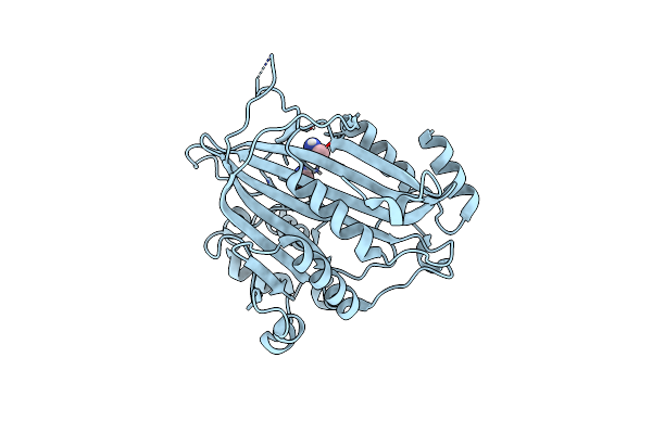
Organism: Synthetic construct, Homo sapiens
Method: X-RAY DIFFRACTION Resolution:2.00 Å Release Date: 2022-01-12 Classification: IMMUNE SYSTEM Ligands: CA, PEG, ACM, NH2 |
 |
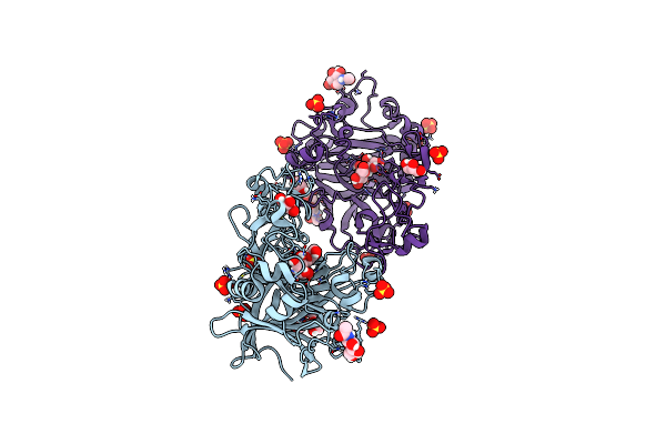
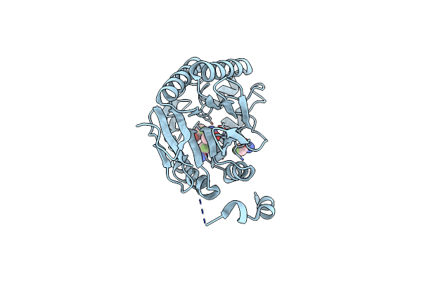
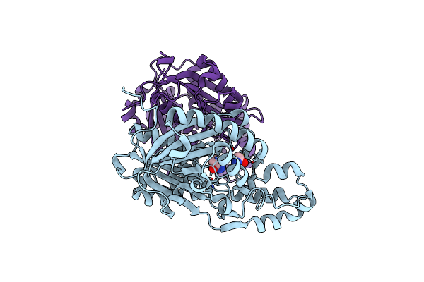
The C146A Variant Of An Amidase From Pyrococcus Horikoshii With Bound Acetamide
Organism: Pyrococcus horikoshii (strain atcc 700860 / dsm 12428 / jcm 9974 / nbrc 100139 / ot-3)
Method: X-RAY DIFFRACTION Resolution:1.65 Å Release Date: 2021-07-21 Classification: HYDROLASE Ligands: ACM, CL |
 |
Organism: Candida albicans (strain sc5314 / atcc mya-2876)
Method: X-RAY DIFFRACTION Resolution:1.58 Å Release Date: 2021-04-21 Classification: TRANSFERASE Ligands: ACM |
 |
Organism: Candida albicans (strain sc5314 / atcc mya-2876)
Method: X-RAY DIFFRACTION Resolution:1.78 Å Release Date: 2021-04-21 Classification: TRANSFERASE Ligands: ACM |
 |
Organism: Humicola insolens
Method: X-RAY DIFFRACTION Resolution:1.88 Å Release Date: 2020-09-16 Classification: HYDROLASE Ligands: NAG, SO4, BGC, YLL, GOL, ACM |
 |
Organism: Humicola insolens
Method: X-RAY DIFFRACTION Resolution:1.20 Å Release Date: 2020-09-16 Classification: HYDROLASE Ligands: GOL, SO4, ACM, NAG |
 |
Organism: Mycobacterium tuberculosis
Method: X-RAY DIFFRACTION Resolution:1.65 Å Release Date: 2020-07-15 Classification: LYASE Ligands: MN, CL, ACM |
 |
Organism: Homo sapiens, Enterobacteria phage t4, Dendroaspis angusticeps
Method: X-RAY DIFFRACTION Resolution:2.55 Å Release Date: 2020-07-08 Classification: MEMBRANE PROTEIN Ligands: ACM, Y01, OIN |
 |
Organism: Akkermansia muciniphila (strain atcc baa-835 / muc)
Method: X-RAY DIFFRACTION Resolution:1.50 Å Release Date: 2019-03-13 Classification: HYDROLASE Ligands: ZN, ACM |
 |
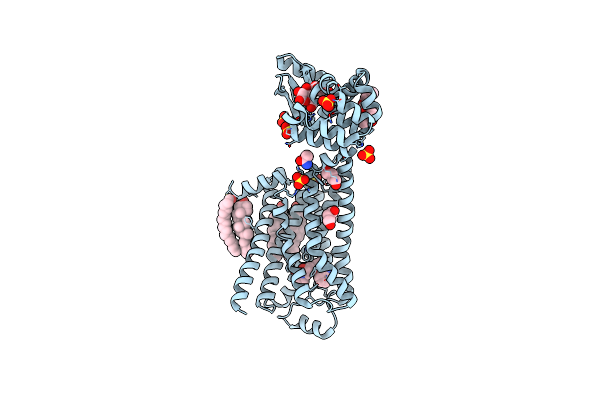
Structure Of Beta2 Adrenoceptor Bound To Carazolol And An Intracellular Allosteric Antagonist
Organism: Homo sapiens, Enterobacteria phage t4
Method: X-RAY DIFFRACTION Resolution:2.70 Å Release Date: 2017-08-16 Classification: SIGNALING PROTEIN Ligands: EPE, CLR, 8VS, CAU, BU1, ACM |
 |
Organism: Homo sapiens, Enterobacteria phage t4
Method: X-RAY DIFFRACTION Resolution:3.20 Å Release Date: 2016-08-17 Classification: MEMBRANE PROTEIN/HYDROLASE Ligands: SO4, CAU, BU1, ACM, CLR, PLM, 12P |
 |
In Meso In Situ Serial X-Ray Crystallography Structure Of The Beta2-Adrenergic Receptor At 100 K
Organism: Homo sapiens, Enterobacteria phage t4
Method: X-RAY DIFFRACTION Resolution:2.48 Å Release Date: 2016-01-13 Classification: Membrane Protein/Hydrolase Ligands: CAU, BU1, ACM, CLR, PLM, 12P, SO4 |
 |
In Meso X-Ray Crystallography Structure Of The Beta2-Adrenergic Receptor At 100 K
Organism: Homo sapiens, Enterobacteria phage t4
Method: X-RAY DIFFRACTION Resolution:3.80 Å Release Date: 2016-01-13 Classification: Membrane Protein/Hydrolase Ligands: CAU, BU1, ACM, CLR, PLM, SO4 |
 |
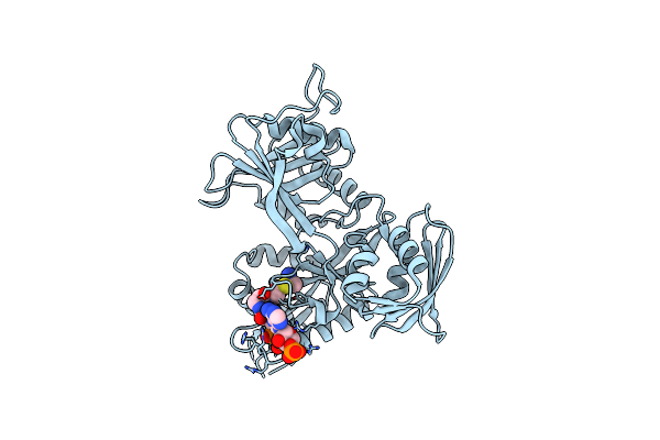
The E41Q Mutant Of The Amidase From Nesterenkonia Sp. An1 Showing Covalent Addition Of The Acetamide Moiety Of Fluoroacetamide At The Active Site Cysteine
Organism: Nesterenkonia sp. 10004
Method: X-RAY DIFFRACTION Resolution:1.92 Å Release Date: 2014-02-12 Classification: hydrolase/substrate Ligands: ACM, FTM |
 |
The E41L Mutant Of The Amidase From Nesterenkonia Sp. An1 Showing Covalent Addition Of The Acetamide Moiety Of Fluoroacetamide At The Active Site Cysteine
Organism: Nesterenkonia sp. 10004
Method: X-RAY DIFFRACTION Resolution:1.60 Å Release Date: 2014-02-12 Classification: HYDROLASE Ligands: ACM, FTM |
 |
Periplasmic Portion Of The Helicobacter Pylori Chemoreceptor Tlpb With Acetamide Bound
Organism: Helicobacter pylori
Method: X-RAY DIFFRACTION Resolution:1.40 Å Release Date: 2012-06-27 Classification: MEMBRANE PROTEIN Ligands: ACM, SO4, GOL |
 |
Organism: Xanthomonas campestris pv. campestris
Method: X-RAY DIFFRACTION Resolution:2.05 Å Release Date: 2012-05-16 Classification: TRANSFERASE Ligands: ACM |
 |
Crystal Structure Of Acetyltransferase Eis From Mycobacterium Tuberculosis H37Rv In Complex With Coa And An Acetamide Moiety
Organism: Mycobacterium tuberculosis
Method: X-RAY DIFFRACTION Resolution:1.95 Å Release Date: 2011-06-01 Classification: TRANSFERASE Ligands: COA, ACM |