
Search Count: 2,852
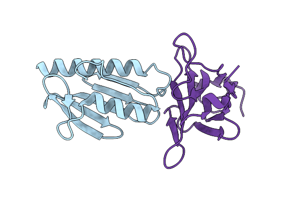 |
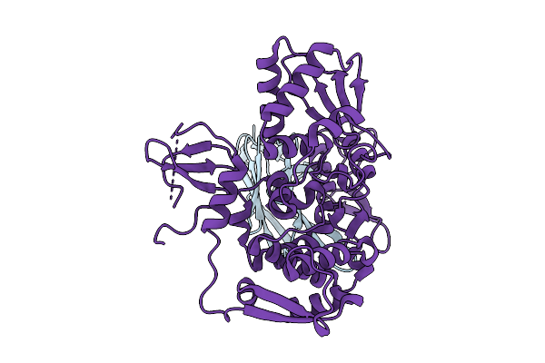
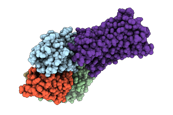
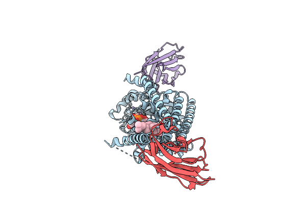
Crystal Structure Of The Human Frataxin Protein In Complex With A Tailored Camelid Nanobody 6B1
Organism: Homo sapiens, Lama glama
Method: X-RAY DIFFRACTION Release Date: 2025-12-24 Classification: OXIDOREDUCTASE |
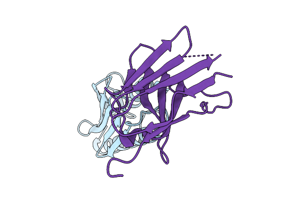 |
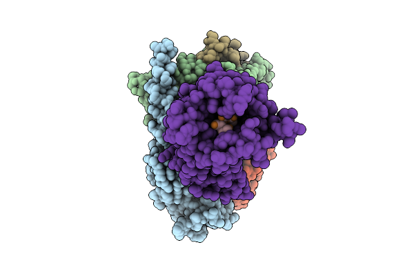
Crystal Structure Of The Human Frataxin Protein In Complex With A Tailored Camelid Nanobody 4A7
Organism: Homo sapiens, Lama glama
Method: X-RAY DIFFRACTION Release Date: 2025-12-24 Classification: OXIDOREDUCTASE |
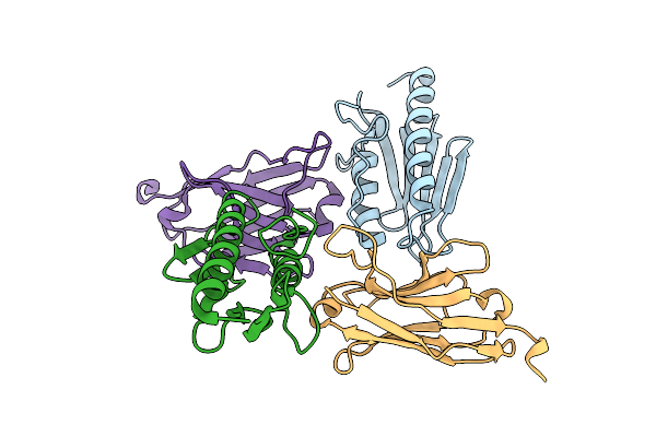 |
Crystal Structure Of The Human Frataxin Protein In Complex With A Tailored Camelid Nanobody 16C10
Organism: Homo sapiens, Lama glama
Method: X-RAY DIFFRACTION Release Date: 2025-12-24 Classification: OXIDOREDUCTASE |
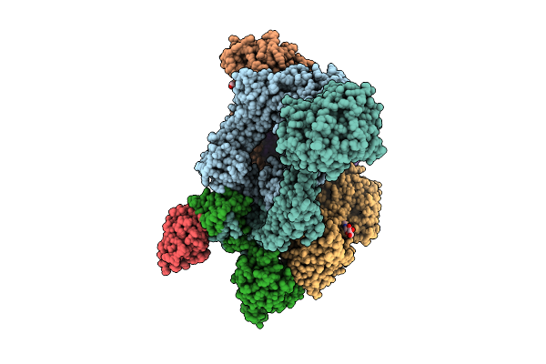 |
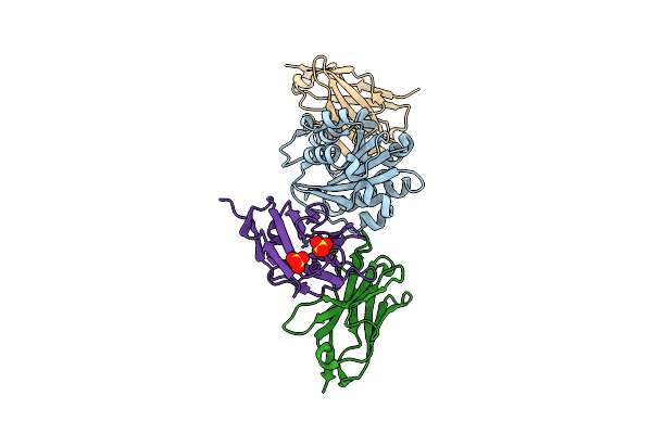
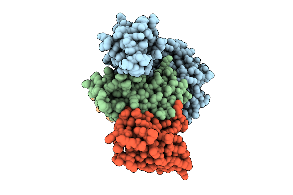
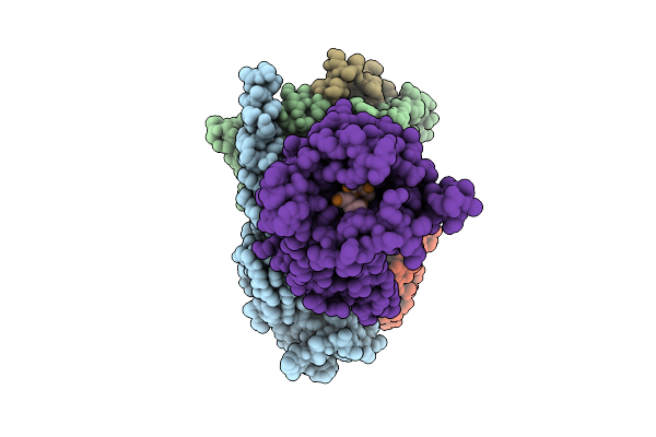
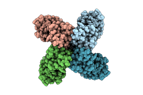
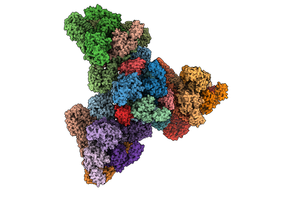
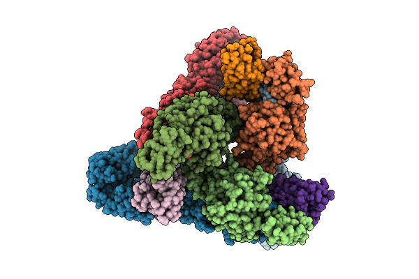
Structure Of The Complement Classical And Lectin Pathway C3 Convertase In Complex With Substrate C3
Organism: Homo sapiens, Lama glama
Method: ELECTRON MICROSCOPY Release Date: 2025-12-24 Classification: IMMUNE SYSTEM Ligands: NAG, MG |
 |
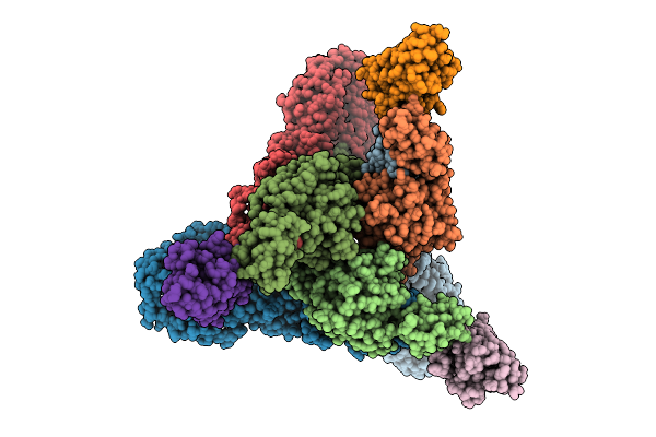
Organism: Lama glama, Homo sapiens
Method: ELECTRON MICROSCOPY Release Date: 2025-12-24 Classification: IMMUNE SYSTEM Ligands: NAG, MG |
 |
Organism: Mus musculus, Lama glama
Method: X-RAY DIFFRACTION Release Date: 2025-12-17 Classification: IMMUNE SYSTEM Ligands: SO4 |
 |
Organism: Homo sapiens, Lama glama, Synthetic construct
Method: ELECTRON MICROSCOPY Release Date: 2025-12-17 Classification: DE NOVO PROTEIN/IMMUNE SYSTEM |
 |
Organism: Lama glama, Homo sapiens
Method: ELECTRON MICROSCOPY Release Date: 2025-12-17 Classification: LIGASE |
 |
Organism: Homo sapiens, Lama glama
Method: ELECTRON MICROSCOPY Release Date: 2025-12-10 Classification: MEMBRANE PROTEIN |
 |
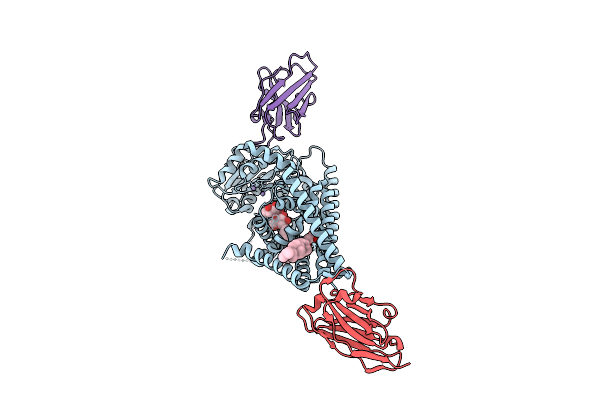
Human Delta Opioid Receptor Complex With Mini-Gi And Agonist Dadle And Allosteric Modulator Mips3614
Organism: Homo sapiens, Lama glama
Method: ELECTRON MICROSCOPY Release Date: 2025-12-10 Classification: MEMBRANE PROTEIN Ligands: A1CVG |
 |
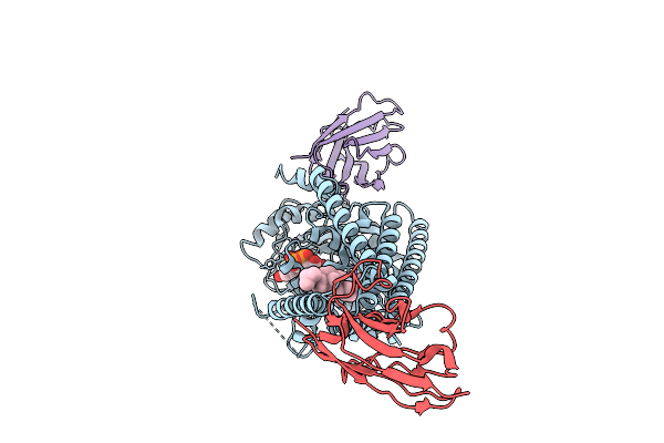
Human Delta Opioid Receptor Complex With Mini-Gi And Agonist Dadle And Allosteric Modulator Mips3983
Organism: Homo sapiens, Lama glama
Method: ELECTRON MICROSCOPY Release Date: 2025-12-10 Classification: MEMBRANE PROTEIN Ligands: A1CU8 |
 |
Organism: Paramecium bursaria chlorella virus cz-2, Lama glama
Method: ELECTRON MICROSCOPY Release Date: 2025-12-10 Classification: TRANSFERASE/IMMUNE SYSTEM Ligands: Y01, MN, LMT |
 |
Organism: Paramecium bursaria chlorella virus cz-2, Lama glama
Method: ELECTRON MICROSCOPY Release Date: 2025-12-10 Classification: TRANSFERASE/IMMUNE SYSTEM Ligands: UGA, MN, Y01 |
 |
Organism: Paramecium bursaria chlorella virus cz-2, Lama glama
Method: ELECTRON MICROSCOPY Release Date: 2025-12-10 Classification: TRANSFERASE/IMMUNE SYSTEM Ligands: UGA, Y01, MN |
 |
Organism: Homo sapiens, Lama glama
Method: ELECTRON MICROSCOPY Release Date: 2025-12-03 Classification: IMMUNE SYSTEM Ligands: K |
 |
Cryo-Em Structure Of [Pen5]-Urotensin (4-11)-Bounded Human Urotensin Receptor (Uts2R)-Gq Complex
Organism: Homo sapiens, Lama glama, Synthetic construct
Method: ELECTRON MICROSCOPY Release Date: 2025-12-03 Classification: SIGNALING PROTEIN Ligands: CHO |
 |
Venezuelan Equine Encephalitis Virus In Complex With The Single Domain Antibody V2B3
Organism: Venezuelan equine encephalitis virus, Lama glama
Method: ELECTRON MICROSCOPY Release Date: 2025-12-03 Classification: Viral protein/Immune System |
 |
Venezuelan Equine Encephalitis Virus In Complex With The Single Domain Antibody V2C3
Organism: Venezuelan equine encephalitis virus, Lama glama
Method: ELECTRON MICROSCOPY Release Date: 2025-12-03 Classification: Viral protein/Immune System |
 |
Venezuelan Equine Encephalitis Virus In Complex With The Single Domain Antibody V3A8F
Organism: Venezuelan equine encephalitis virus, Lama glama
Method: ELECTRON MICROSCOPY Release Date: 2025-12-03 Classification: Viral protein/Immune System |
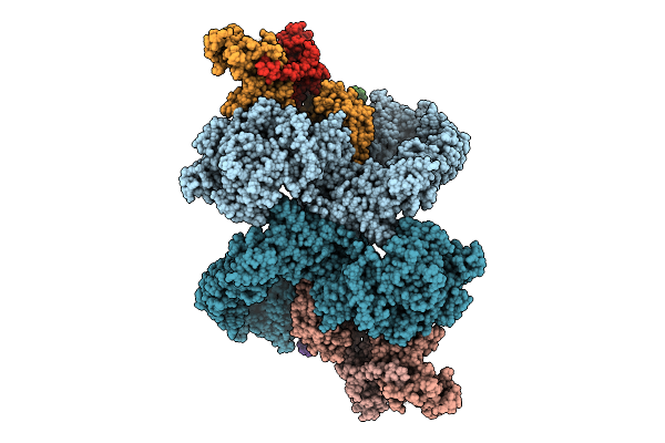 |
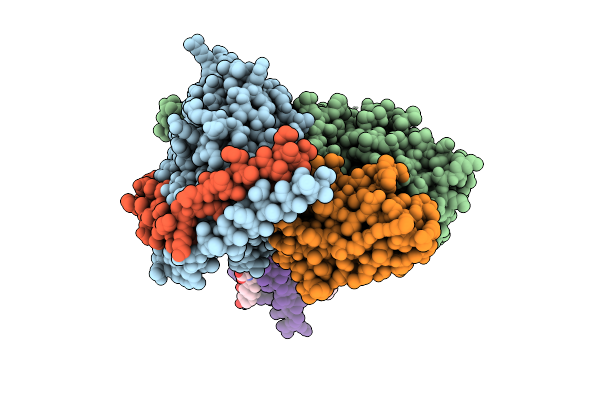
Cryo-Em Structure Of The Pi4Ka Complex Bound To An Efr3 Interfering Nanobody (F3In)
Organism: Homo sapiens, Lama glama
Method: ELECTRON MICROSCOPY Release Date: 2025-12-03 Classification: SIGNALING PROTEIN |