
Search Count: 1,28,006
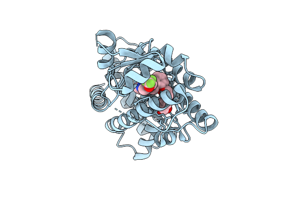 |
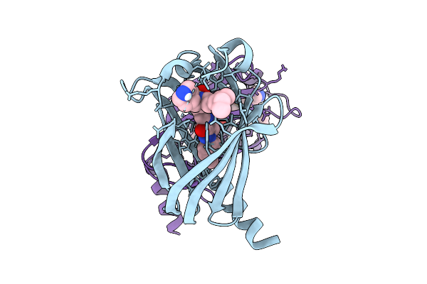
X-Ray Structure Of Human Ppar Delta Ligand Binding Domain In Complex With A Synthetic Agonist 16A
Organism: Homo sapiens
Method: X-RAY DIFFRACTION Release Date: 2025-12-24 Classification: NUCLEAR PROTEIN Ligands: A1D86 |
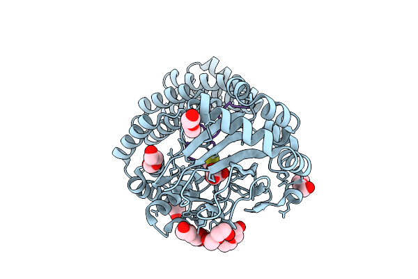 |
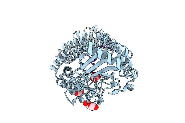
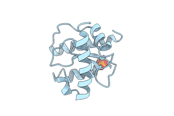
Aspartyl/Asparaginyl Beta-Hydroxylase (Asph) In Complex With Fe, 2-Oxoglutarate, Thiocyanate And Factor X Derived Peptide Fragment
Organism: Homo sapiens
Method: X-RAY DIFFRACTION Release Date: 2025-12-24 Classification: OXIDOREDUCTASE Ligands: AKG, FE, PEG, SCN |
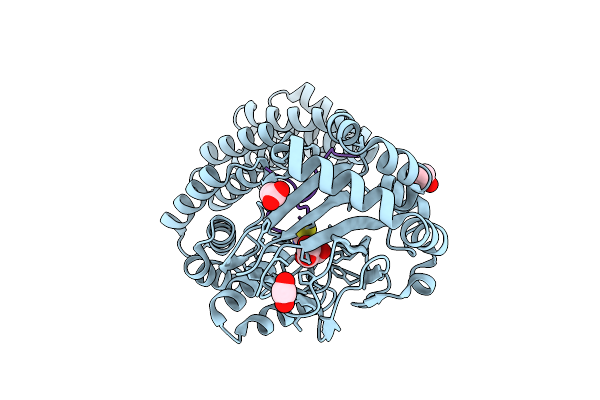 |
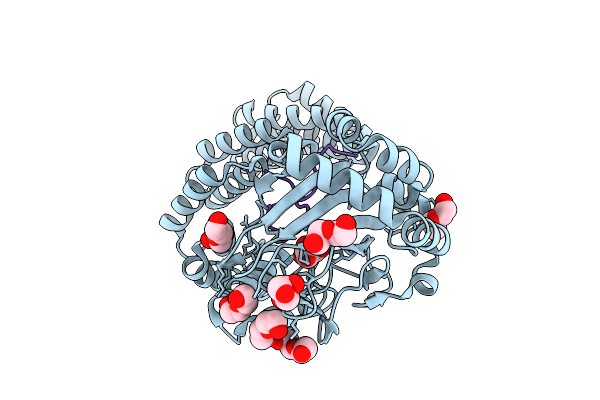
Aspartyl/Asparaginyl Beta-Hydroxylase (Asph) In Complex With Mn, 2Og, Thiocyanate And Factor X Peptide Fragment (39Mer-4Ser)
Organism: Homo sapiens
Method: X-RAY DIFFRACTION Release Date: 2025-12-24 Classification: OXIDOREDUCTASE Ligands: AKG, ACT, PEG, MN, SCN |
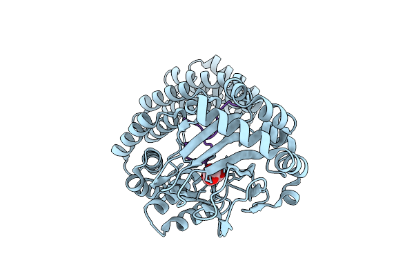 |
Aspartyl/Asparaginyl Beta-Hydroxylase (Asph) In Complex With Fe, 2Og, Sin And Hydroxylated Product Of Factor X Peptide Fragment (39Mer-4Ser)
Organism: Homo sapiens
Method: X-RAY DIFFRACTION Release Date: 2025-12-24 Classification: OXIDOREDUCTASE Ligands: SIN, AKG, FE |
 |
Aspartyl/Asparaginyl Beta-Hydroxylase (Asph) In Complex With Fe, 2-Oxoglutarate And Factor X Derived Peptide Fragment
Organism: Homo sapiens
Method: X-RAY DIFFRACTION Release Date: 2025-12-24 Classification: OXIDOREDUCTASE Ligands: AKG, PEG, CL, FE |
 |
Aspartyl/Asparaginyl Beta-Hydroxylase (Asph) In Complex With Fe, 2-Oxoglutarate, Succinate And The Hudroxylated Product Of Factor X Derived Peptide Fragment After O2 Exposure
Organism: Homo sapiens
Method: X-RAY DIFFRACTION Release Date: 2025-12-24 Classification: OXIDOREDUCTASE Ligands: AKG, SIN, PEG, FE, GOL, CL |
 |
Aspartyl/Asparaginyl Beta-Hydroxylase (Asph) In Complex With Fe, 2-Oxoglutarate And A Factor X Derived Peptide Fragment
Organism: Homo sapiens
Method: X-RAY DIFFRACTION Release Date: 2025-12-24 Classification: OXIDOREDUCTASE Ligands: AKG, PEG, GOL, FE |
 |
Room Temperature Structure Of Aspartyl/Asparaginyl Beta-Hydroxylase (Asph) In Complex With Fe, 2-Oxoglutarate And Factor X Derived Peptide Fragment
Organism: Homo sapiens
Method: X-RAY DIFFRACTION Release Date: 2025-12-24 Classification: OXIDOREDUCTASE Ligands: AKG, FE |
 |
Organism: Homo sapiens
Method: X-RAY DIFFRACTION Release Date: 2025-12-24 Classification: LIPID BINDING PROTEIN Ligands: A1IWC, GOL, PEG |
 |
Organism: Homo sapiens
Method: X-RAY DIFFRACTION Release Date: 2025-12-24 Classification: LIPID BINDING PROTEIN Ligands: A1IWD |
 |
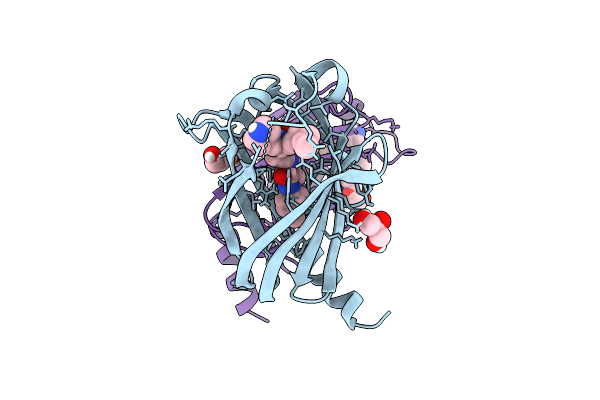
X-Ray Structure Of The C-Terminal Domain (Residues 366-485) Of S. Pombe Threonylcarbamoyladenosine Dehydratase
Organism: Schizosaccharomyces pombe
Method: X-RAY DIFFRACTION Release Date: 2025-12-24 Classification: RNA BINDING PROTEIN Ligands: PO4 |
 |
Organism: Homo sapiens
Method: X-RAY DIFFRACTION Release Date: 2025-12-24 Classification: HYDROLASE Ligands: GDP, EDO, A1IWE |
 |
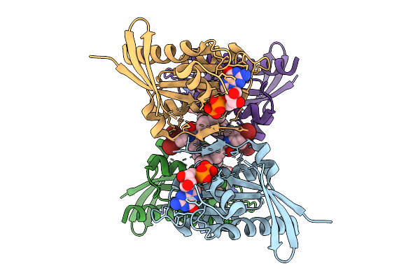
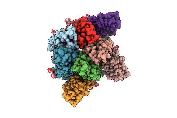
A5B3 Gabaa Receptor Bound To Gaba And Mb25 In Desensitized State In Detergent Micelles
Organism: Aequorea victoria, Homo sapiens, Escherichia coli
Method: ELECTRON MICROSCOPY Release Date: 2025-12-24 Classification: MEMBRANE PROTEIN Ligands: ABU, EPE, NAG |
 |
Organism: Aequorea victoria, Homo sapiens, Escherichia coli
Method: ELECTRON MICROSCOPY Release Date: 2025-12-24 Classification: MEMBRANE PROTEIN Ligands: ABU, EPE |
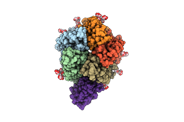 |
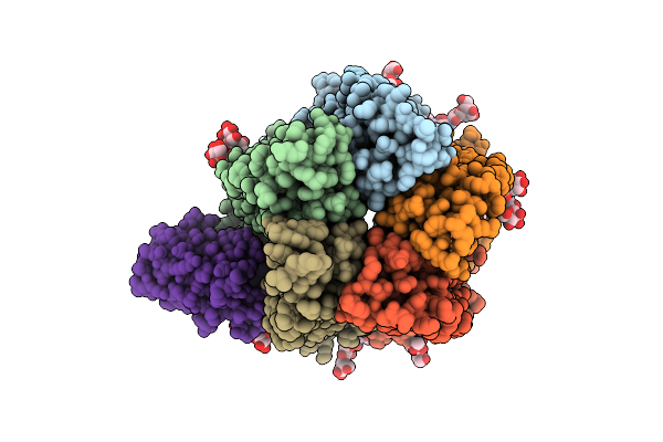
A5B3 Gabaar Bound To Gaba And Mb25 In A Desensitized State In Saposin Nanodiscs After Long Gaba Treatment
Organism: Aequorea victoria, Homo sapiens, Escherichia coli
Method: ELECTRON MICROSCOPY Release Date: 2025-12-24 Classification: MEMBRANE PROTEIN Ligands: ABU, EPE, NAG |
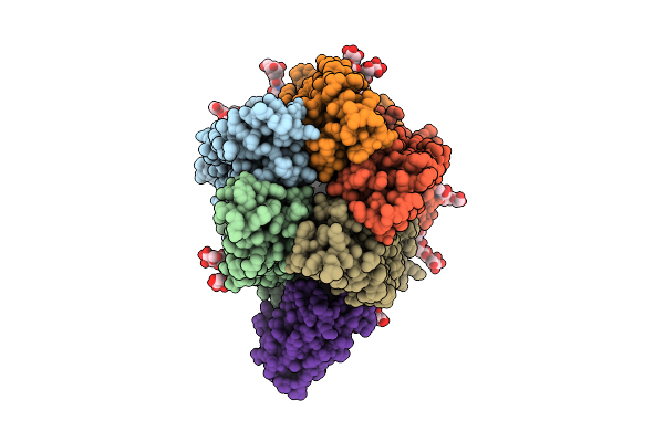 |
A5B3 Gabaar Bound To Etomidate, Gaba, And Mb25 In A Desensitized State In Saposin Nanodiscs
Organism: Aequorea victoria, Homo sapiens, Escherichia coli
Method: ELECTRON MICROSCOPY Release Date: 2025-12-24 Classification: MEMBRANE PROTEIN Ligands: V8D, ABU, EPE, NAG |
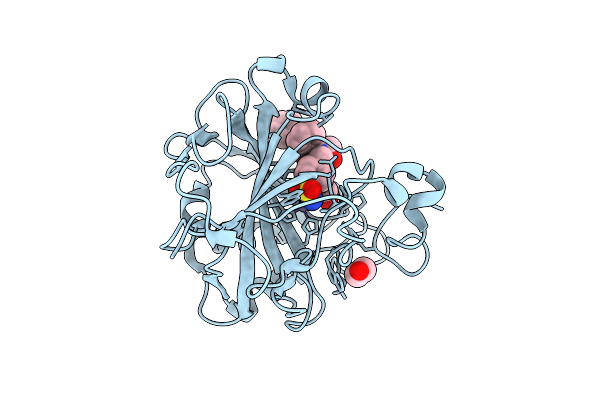 |
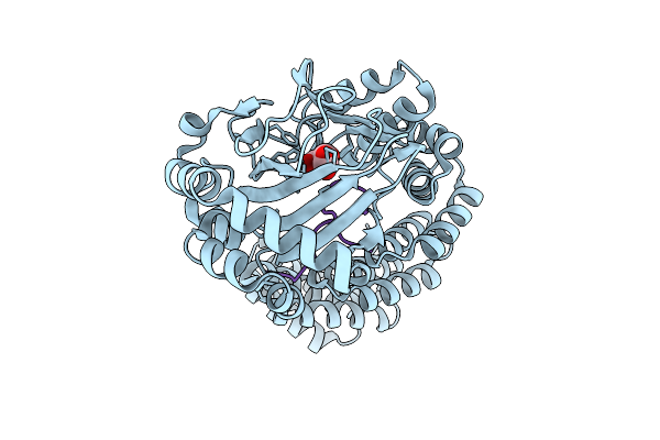
Crystal Structure Of Human Carbonic Anhydrase Ii In Complex With N-Benzyl-2-(2-Chloro-N-(4-Sulfamoylphenethyl)Acetamido)-2-Phenylacetamide
Organism: Homo sapiens
Method: X-RAY DIFFRACTION Release Date: 2025-12-24 Classification: LYASE Ligands: GOL, EDO, ZN, A1IWH |
 |
Room Temperature Structure Of Aspartyl/Asparaginyl Beta-Hydroxylase (Asph) In Complex With Fe, 2-Oxoglutarate And Hydroxylated Factor X Derived Peptide Fragment, 2 H O2 Exposure
Organism: Homo sapiens
Method: X-RAY DIFFRACTION Release Date: 2025-12-24 Classification: OXIDOREDUCTASE Ligands: SIN, FE |
 |
Aspartyl/Asparaginyl Beta-Hydroxylase (Asph) In Complex With Fe, 2-Oxoglutarate, Succinate And The Hydroxylated Product Of Factor X Derived Peptide Fragment, 12 H O2 Exposure
Organism: Homo sapiens
Method: X-RAY DIFFRACTION Release Date: 2025-12-24 Classification: OXIDOREDUCTASE Ligands: SIN, FE |
 |
Room Temperature Structure Of Aspartyl/Asparaginyl Beta-Hydroxylase (Asph) In Complex With Fe, 2-Oxoglutarate, And Hydroxylated Factor X Derived Peptide Fragment, 1.5 S O2 Exposure
Organism: Homo sapiens
Method: X-RAY DIFFRACTION Release Date: 2025-12-24 Classification: OXIDOREDUCTASE Ligands: AKG, SIN, FE |