
Search Count: 1,901
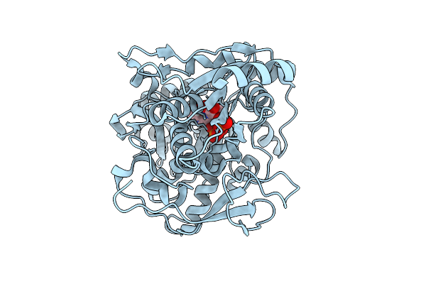 |
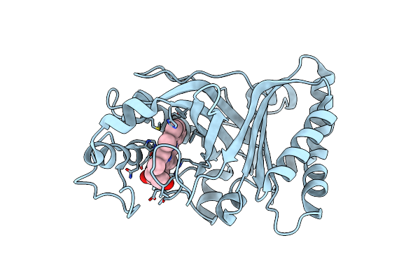
Crystal Structure Of Capa2 From Alistipes Finegoldii In Complex With Ops-Plp External Aldimine
Organism: Alistipes finegoldii dsm 17242
Method: X-RAY DIFFRACTION Release Date: 2025-11-05 Classification: BIOSYNTHETIC PROTEIN Ligands: E1U |
 |
Organism: Actinoalloteichus caeruleus
Method: X-RAY DIFFRACTION Release Date: 2025-10-22 Classification: BIOSYNTHETIC PROTEIN Ligands: SAH |
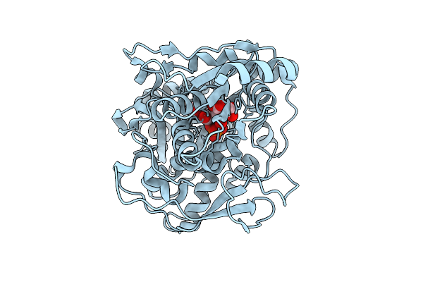 |
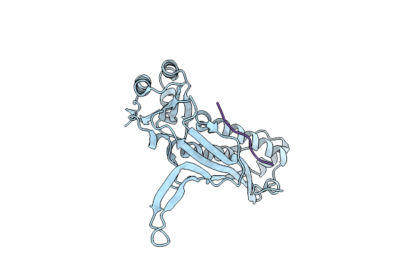
Crystal Structure Of Capa2 From Alistipes Finegoldii In Complex With Plp Cofactor
Organism: Alistipes finegoldii dsm 17242
Method: X-RAY DIFFRACTION Release Date: 2025-10-22 Classification: BIOSYNTHETIC PROTEIN Ligands: CIT, PLP |
 |
Organism: Ostrinia furnacalis
Method: SOLID-STATE NMR Release Date: 2025-10-22 Classification: STRUCTURAL PROTEIN |
 |
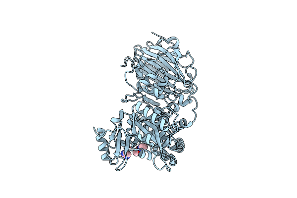
Crystal Structure Of The Catalytic Domain From The Bont-Like Toxin Complex Pg1 Of Paeniclostridium Ghonii
Organism: Paraclostridium ghonii
Method: X-RAY DIFFRACTION Release Date: 2025-10-15 Classification: TOXIN Ligands: ZN, SO4, ACT, GOL |
 |
Organism: Hbv genotype d3, Homo sapiens
Method: ELECTRON MICROSCOPY Release Date: 2025-07-02 Classification: VIRAL PROTEIN/IMMUNE SYSTEM |
 |
Organism: Homo sapiens, Hbv genotype d3
Method: ELECTRON MICROSCOPY Release Date: 2025-07-02 Classification: IMMUNE SYSTEM/VIRAL PROTEIN |
 |
Organism: Streptomyces sp. sn-593
Method: X-RAY DIFFRACTION Release Date: 2025-07-02 Classification: OXIDOREDUCTASE Ligands: HEM, RRM, CL |
 |
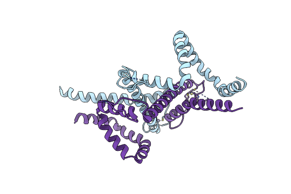
Cryo-Em Structure And Molecular Assembly Of The Bont-Like Toxin Pg1 Complex From Paeniclostridium Ghonii
Organism: Paraclostridium ghonii
Method: ELECTRON MICROSCOPY Resolution:3.20 Å Release Date: 2025-03-26 Classification: TOXIN |
 |
Organism: Escherichia coli
Method: ELECTRON MICROSCOPY Release Date: 2025-03-19 Classification: OXIDOREDUCTASE Ligands: HEM |
 |
Organism: Escherichia coli
Method: ELECTRON MICROSCOPY Release Date: 2025-03-19 Classification: VIRUS LIKE PARTICLE |
 |
Organism: Chromobacterium haemolyticum
Method: X-RAY DIFFRACTION Resolution:1.35 Å Release Date: 2025-01-29 Classification: TOXIN Ligands: IMD, ACT |
 |
Structure Of Hbv Surface Antigen Determined In Recombinant Spherical Subviral Particle
Organism: Hbv genotype d3
Method: ELECTRON MICROSCOPY Release Date: 2025-01-15 Classification: VIRAL PROTEIN Ligands: ZN |
 |
The Structure Of Type Iii Crispr-Associated Deaminase In Complex Ca6 And Atp, Fully Activated
Organism: Limisphaera ngatamarikiensis
Method: ELECTRON MICROSCOPY Resolution:3.40 Å Release Date: 2024-12-25 Classification: IMMUNE SYSTEM Ligands: ATP, ZN, MG |
 |
Organism: Limisphaera ngatamarikiensis
Method: ELECTRON MICROSCOPY Release Date: 2024-12-25 Classification: IMMUNE SYSTEM Ligands: ZN, LQJ |
 |
Organism: Limisphaera ngatamarikiensis
Method: ELECTRON MICROSCOPY Release Date: 2024-12-11 Classification: IMMUNE SYSTEM Ligands: ZN, ATP |
 |
Organism: Limisphaera ngatamarikiensis
Method: ELECTRON MICROSCOPY Release Date: 2024-12-11 Classification: IMMUNE SYSTEM |
 |
High-Resolution Structure Of The Siderophore Periplasmic Binding Protein Ftsb From Streptococcus Pyogenes
Organism: Streptococcus pyogenes ssi-1
Method: X-RAY DIFFRACTION Resolution:1.11 Å Release Date: 2024-10-09 Classification: METAL BINDING PROTEIN Ligands: P33, PEG, EDO, P6G, ZN, NA, CL |
 |
Structure Of The Siderophore Periplasmic Binding Protein Ftsb From Streptococcus Pyogenes With Ferrichrome Bound
Organism: Streptococcus pyogenes ssi-1
Method: X-RAY DIFFRACTION Resolution:1.80 Å Release Date: 2024-10-09 Classification: METAL BINDING PROTEIN Ligands: FCE |
 |
Structure Of The Siderophore Periplasmic Binding Protein Ftsb From Streptococcus Pyogenes With Bisucaberin Bound
Organism: Streptococcus pyogenes ssi-1
Method: X-RAY DIFFRACTION Resolution:2.00 Å Release Date: 2024-10-09 Classification: METAL BINDING PROTEIN Ligands: OX8, FE |