Planned Maintenance: Some services may turn out to be unavailable from 15th January, 2026 to 16th January, 2026. We apologize for the inconvenience!

Planned Maintenance: Some services may turn out to be unavailable from 15th January, 2026 to 16th January, 2026. We apologize for the inconvenience!
 |
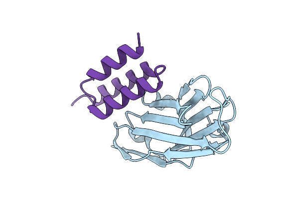
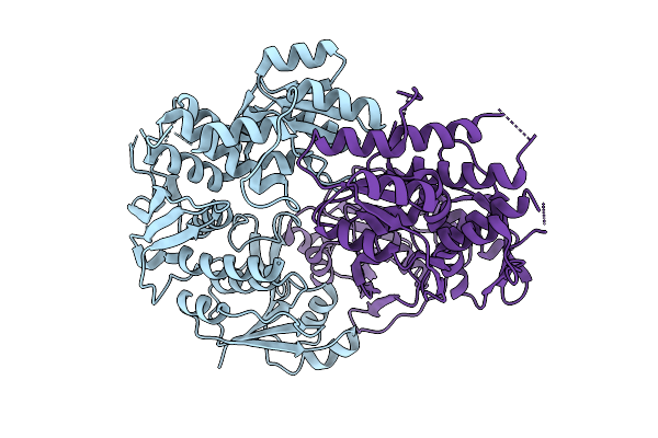
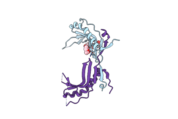
Crystal Structure Of S. Aureus Protein A Bound To A Human Single-Domain Antibody
Organism: Homo sapiens, Staphylococcus aureus (strain nctc 8325 / ps 47)
Method: X-RAY DIFFRACTION Release Date: 2025-11-12 Classification: IMMUNE SYSTEM |
 |
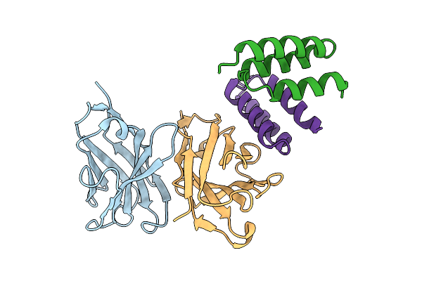
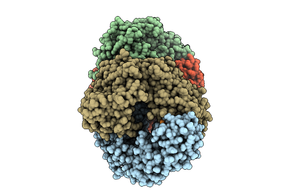
Crystal Structure Of S. Aureus Protein A Bound To A Camelid Single-Domain Antibody
Organism: Camelidae, Staphylococcus aureus subsp. aureus nctc 8325
Method: X-RAY DIFFRACTION Release Date: 2025-11-12 Classification: IMMUNE SYSTEM |
 |
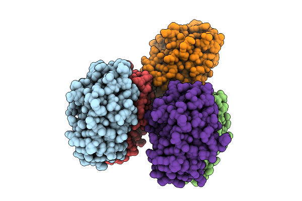
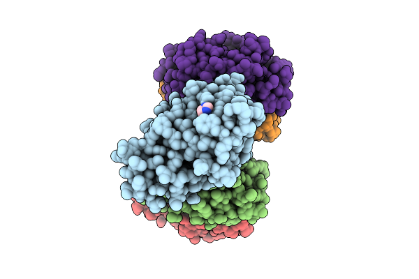
Crystal Structure Of S. Aureus Protein A Bound To A Camelid Single-Domain Antibody
Organism: Staphylococcus aureus (strain nctc 8325 / ps 47), Camelus dromedarius
Method: X-RAY DIFFRACTION Release Date: 2025-11-12 Classification: IMMUNE SYSTEM |
 |
Organism: Glutamicibacter protophormiae
Method: ELECTRON MICROSCOPY Release Date: 2025-10-15 Classification: HYDROLASE Ligands: CA, DUC |
 |
Crystal Structure Of A Phgs Rhamnosyltransferase Ugt79G15 From Rehmannia Glutinosa
Organism: Rehmannia glutinosa
Method: X-RAY DIFFRACTION Release Date: 2025-10-08 Classification: TRANSFERASE |
 |
Crystal Structure Of A Phgs Rhamnosyltransferase Ugt79G15 From Rehmannia Glutinosa In Complex With Udp
Organism: Rehmannia glutinosa
Method: X-RAY DIFFRACTION Release Date: 2025-10-08 Classification: TRANSFERASE Ligands: UDP |
 |
Crystal Structure Of A Phgs Rhamnosyltransferase Ugt79G15 From Rehmannia Glutinosa In Complex With Udp And Fsa
Organism: Rehmannia glutinosa
Method: X-RAY DIFFRACTION Release Date: 2025-10-08 Classification: TRANSFERASE Ligands: A1EO9, UDP |
 |
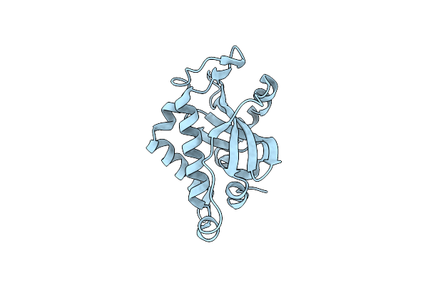
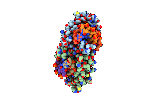
Solution Structure Of Staphylococcus Aureus Response Regulator Arlr Dna-Binding Domain
Organism: Staphylococcus aureus subsp. aureus nctc 8325
Method: SOLUTION NMR Release Date: 2025-09-17 Classification: DNA BINDING PROTEIN |
 |
Organism: Pseudomonas brassicacearum
Method: X-RAY DIFFRACTION Release Date: 2025-07-23 Classification: BIOSYNTHETIC PROTEIN Ligands: IMD |
 |
Organism: Sulfolobus islandicus rey15a
Method: X-RAY DIFFRACTION Release Date: 2025-07-23 Classification: IMMUNE SYSTEM |
 |
Organism: Saccharolobus islandicus rey15a, Synthetic construct
Method: X-RAY DIFFRACTION Release Date: 2025-07-23 Classification: IMMUNE SYSTEM/RNA |
 |
Organism: Pseudomonas brassicacearum
Method: X-RAY DIFFRACTION Release Date: 2025-07-23 Classification: BIOSYNTHETIC PROTEIN Ligands: IMD |
 |
Crystal Structure Of The Tin2-Fold Effector Protein Tue1 From Thecaphora Thlaspeos
Organism: Thecaphora thlaspeos
Method: X-RAY DIFFRACTION Release Date: 2025-06-25 Classification: NUCLEAR PROTEIN |
 |
Crystal Structure Of Staphylococcus Aureus Ding Protein In Complex With Ssdna
Organism: Staphylococcus aureus (strain nctc 8325 / ps 47), Escherichia coli
Method: X-RAY DIFFRACTION Release Date: 2025-06-25 Classification: HYDROLASE/DNA |
 |
Crystal Structure In Space Group C2221 Of A Nucleoid-Associated Protein (Ubp) From Sulfolobus Islandicus.
Organism: Sulfolobus islandicus rey15a
Method: X-RAY DIFFRACTION Release Date: 2025-06-11 Classification: DNA BINDING PROTEIN Ligands: PEG, PG4 |
 |
Crystal Structure In Space Group P21 Of A Nucleoid-Associated Protein (Ubp) From Sulfolobus Islandicus.
Organism: Sulfolobus islandicus rey15a
Method: X-RAY DIFFRACTION Release Date: 2025-06-11 Classification: DNA BINDING PROTEIN Ligands: PEG, EDO |
 |
Crystal Structure Of A Nucleoid-Associated Protein (Ubp) Bound To Dna From Sulfolobus Islandicus.
Organism: Sulfolobus islandicus rey15a
Method: X-RAY DIFFRACTION Release Date: 2025-06-11 Classification: DNA BINDING PROTEIN |
 |
Crystal Structure Of Staphylococcus Aureus Ding Protein In Complex With Ssdna And Ca2+
Organism: Staphylococcus aureus (strain nctc 8325 / ps 47), Chemical production metagenome
Method: X-RAY DIFFRACTION Resolution:3.21 Å Release Date: 2025-05-21 Classification: HYDROLASE/DNA Ligands: CA |
 |
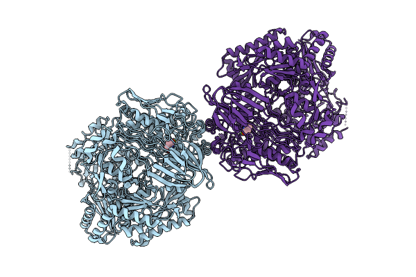
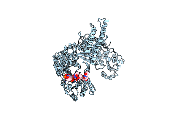
Crystal Structure Of Bifunctional Glmu From Staphylococcus Aureus Nctc 8325 Complexed With Acetyl Coa And Citrate
Organism: Staphylococcus aureus subsp. aureus nctc 8325
Method: X-RAY DIFFRACTION Resolution:1.90 Å Release Date: 2025-05-14 Classification: TRANSFERASE Ligands: ACO, CIT |
 |
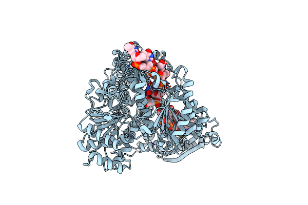
Crystal Structure Of Bifunctional Glmu From Staphylococcus Aureus Nctc 8325 Complexed With Utp, Coa And Glc 1-P
Organism: Staphylococcus aureus subsp. aureus nctc 8325
Method: X-RAY DIFFRACTION Resolution:1.85 Å Release Date: 2025-05-14 Classification: TRANSFERASE Ligands: COA, MG, G1P, UTP |