
Search Count: 243
 |
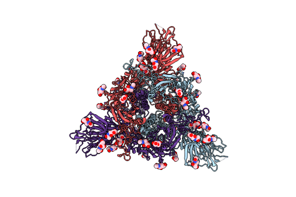
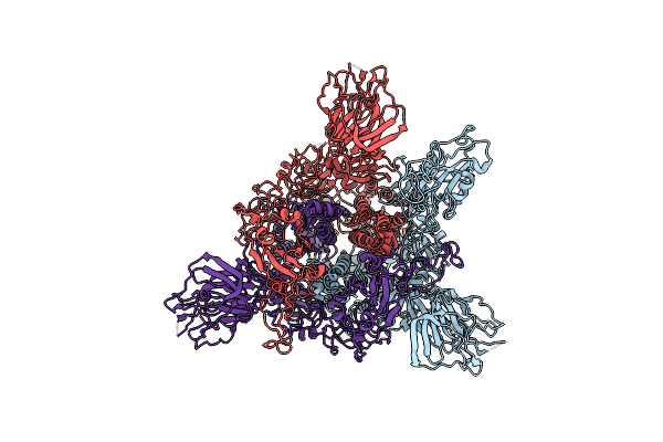
Crystal Structure Of Gamma-Glutamyl-Methylamide Synthetase From Methylovorus Mays (Mmgmas) In Complex With Atpgs
Organism: Methylovorus mays
Method: X-RAY DIFFRACTION Resolution:2.65 Å Release Date: 2026-01-28 Classification: LIGASE Ligands: AGS, MG |
 |
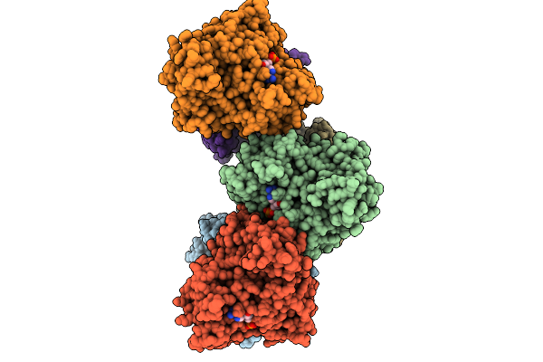
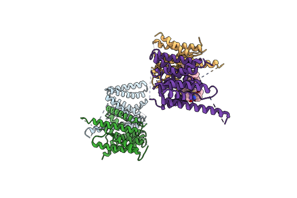
Structure Of Vsd4-Nav1.7-Navpas Channel Chimera Bound To The Hybrid Inhibitor Gne-9296
Organism: Homo sapiens, Periplaneta americana, Diguetia canities
Method: ELECTRON MICROSCOPY Release Date: 2023-04-12 Classification: MEMBRANE PROTEIN/INHIBITOR Ligands: NAG, X80, PEE, Y01 |
 |
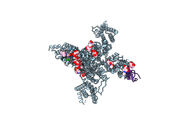
Complex Of Voltage-Gated Sodium Channel Navpas From American Cockroach Periplaneta Americana And Dc1A
Organism: Periplaneta americana, Diguetia canities
Method: ELECTRON MICROSCOPY Resolution:2.80 Å Release Date: 2018-08-08 Classification: MEMBRANE PROTEIN/TOXIN Ligands: NAG, NA |
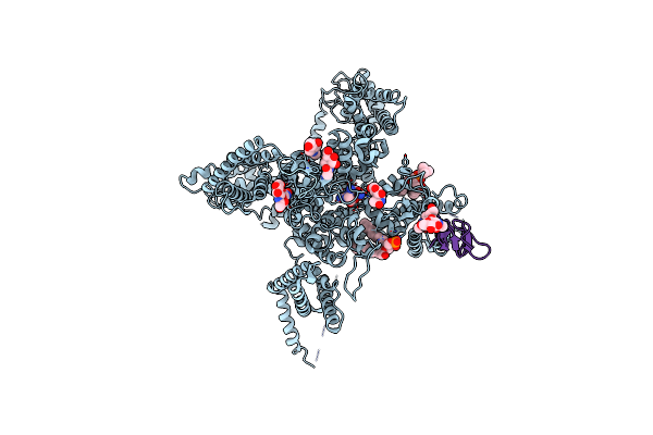 |
Complex Of Voltage-Gated Sodium Channel Navpas From American Cockroach Periplaneta Americana Bound With Saxitoxin And Dc1A
Organism: Periplaneta americana, Diguetia canities
Method: ELECTRON MICROSCOPY Resolution:3.20 Å Release Date: 2018-08-08 Classification: MEMBRANE PROTEIN/TOXIN Ligands: NAG, PC1, 9SL |
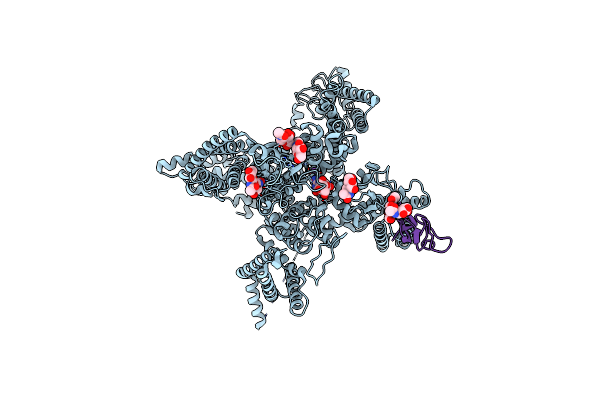 |
Complex Of Voltage-Gated Sodium Channel Navpas From American Cockroach Periplaneta Americana Bound With Tetrodotoxin And Dc1A
Organism: Periplaneta americana, Diguetia canities
Method: ELECTRON MICROSCOPY Resolution:2.60 Å Release Date: 2018-08-08 Classification: MEMBRANE PROTEIN/TOXIN Ligands: NAG, 9SR, NA |
 |
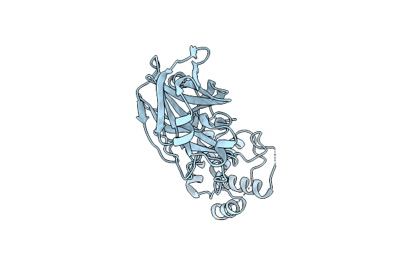
Organism: Sars coronavirus bj01
Method: ELECTRON MICROSCOPY Release Date: 2017-05-03 Classification: VIRAL PROTEIN Ligands: NAG |
 |
Organism: Sars coronavirus bj01
Method: ELECTRON MICROSCOPY Release Date: 2017-05-03 Classification: VIRAL PROTEIN |
 |
Organism: Proteus myxofaciens
Method: X-RAY DIFFRACTION Resolution:2.00 Å Release Date: 2016-04-06 Classification: HYDROLASE Ligands: FAD |
 |
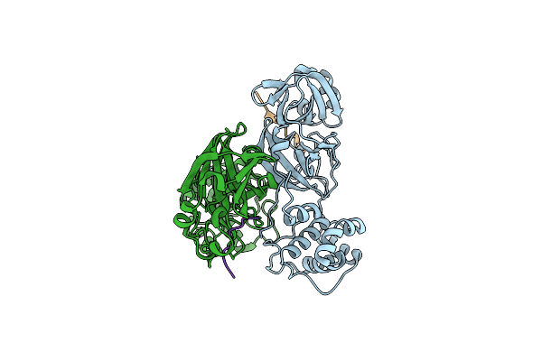
Structure Of L-Amino Acid Deaminase From Proteus Myxofaciens In Complex With Anthranilate
Organism: Proteus myxofaciens
Method: X-RAY DIFFRACTION Resolution:1.75 Å Release Date: 2016-04-06 Classification: HYDROLASE Ligands: FAD, BE2 |
 |
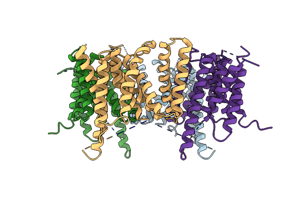
Organism: Methanoculleus marisnigri jr1
Method: X-RAY DIFFRACTION Resolution:3.86 Å Release Date: 2015-03-18 Classification: MEMBRANE PROTEIN/INHIBITOR Ligands: 4B5 |
 |
 |
Nmr Structure Of Temporin-1 Ta In Lipopolysaccharide Micelles: Mechanistic Insight Into Inactivation By Outer Memebrane
Organism: Rana temporaria
Method: SOLUTION NMR Release Date: 2013-08-21 Classification: IMMUNE SYSTEM |
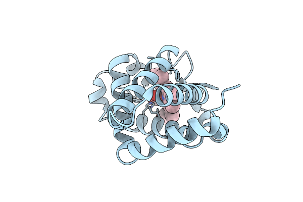 |
Crystal Structure Of The Neuroglobin From The Photosymbiotic Marine Acoel Symsagittifera Roscoffensis
Organism: Symsagittifera roscoffensis
Method: X-RAY DIFFRACTION Resolution:2.30 Å Release Date: 2013-01-09 Classification: TRANSPORT PROTEIN Ligands: HEM, OXY |
 |
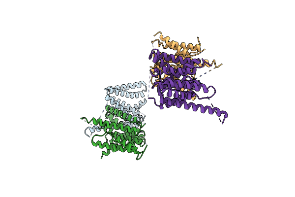
Structure Of A Presenilin Family Intramembrane Aspartate Protease In P2 Space Group
Organism: Methanoculleus marisnigri jr1
Method: X-RAY DIFFRACTION Resolution:3.95 Å Release Date: 2012-12-19 Classification: MEMBRANE PROTEIN |
 |
Structure Of A Presenilin Family Intramembrane Aspartate Protease In C2221 Space Group
Organism: Methanoculleus marisnigri jr1
Method: X-RAY DIFFRACTION Resolution:3.80 Å Release Date: 2012-12-19 Classification: MEMBRANE PROTEIN |
 |
Structure Of A Presenilin Family Intramembrane Aspartate Protease In C222 Space Group
Organism: Methanoculleus marisnigri jr1
Method: X-RAY DIFFRACTION Resolution:3.32 Å Release Date: 2012-12-19 Classification: MEMBRANE PROTEIN |
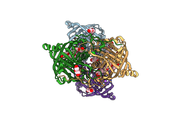 |
Crystal Structure Of Acetoacetate Decarboxylase (Yp_001047042.1) From Methanoculleus Marisnigri Jr1 At 1.60 A Resolution
Organism: Methanoculleus marisnigri jr1
Method: X-RAY DIFFRACTION Resolution:1.60 Å Release Date: 2008-04-01 Classification: LYASE |
 |
Crystal Structure Of Sars-Cov Main Protease H41A Mutant In Complex With An N-Terminal Substrate
Organism: Sars coronavirus
Method: X-RAY DIFFRACTION Resolution:2.50 Å Release Date: 2008-02-12 Classification: HYDROLASE |
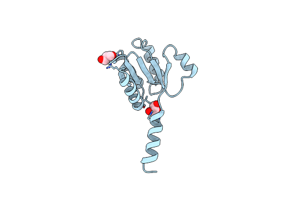 |
Crystal Structure Of The N-Terminal Domain Of Response Regulator Receiver Protein From Methanoculleus Marisnigri Jr1
Organism: Methanoculleus marisnigri jr1
Method: X-RAY DIFFRACTION Resolution:1.70 Å Release Date: 2008-02-05 Classification: SIGNALING PROTEIN Ligands: GOL |
 |
Organism: Sars coronavirus
Method: X-RAY DIFFRACTION Resolution:2.50 Å Release Date: 2007-10-30 Classification: HYDROLASE |