
Search Count: 2,000
 |
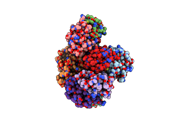
Organism: Mycobacterium tuberculosis (strain cdc 1551 / oshkosh)
Method: X-RAY DIFFRACTION Release Date: 2025-06-11 Classification: TOXIN |
 |
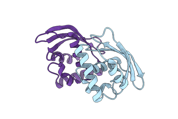
Organism: Mycobacterium tuberculosis
Method: X-RAY DIFFRACTION Release Date: 2025-06-11 Classification: TOXIN |
 |
Organism: Mycobacterium tuberculosis cdc1551
Method: X-RAY DIFFRACTION Resolution:2.68 Å Release Date: 2024-05-29 Classification: IMMUNE SYSTEM |
 |
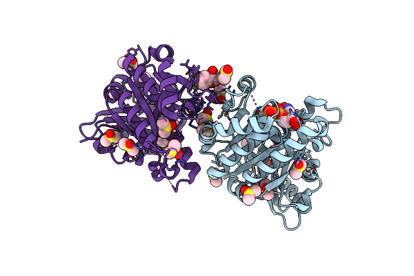
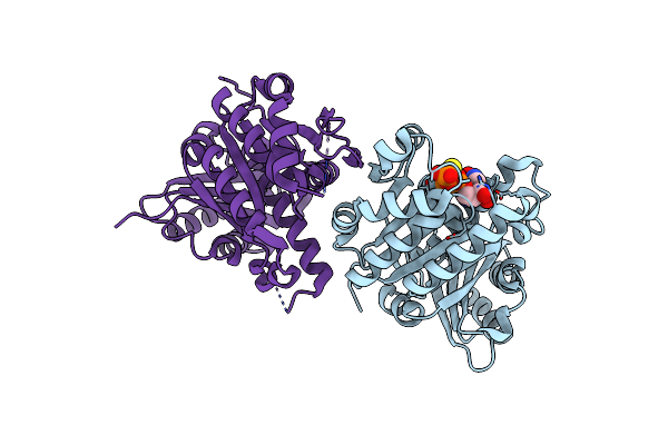
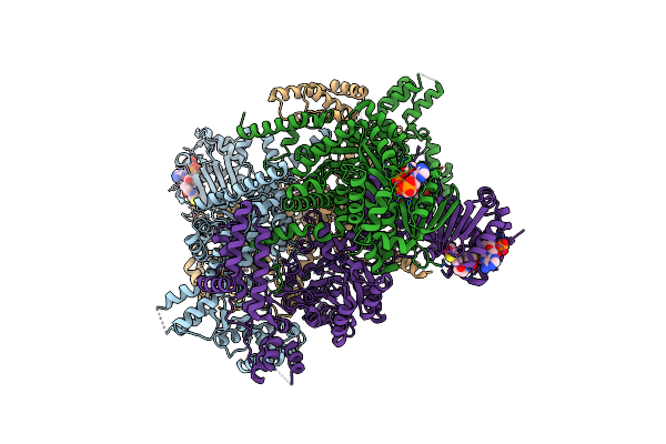
Structural Insights Into Mycobacterium Tuberculosis Clpp1P2 Inhibition By Cediranib: Implications For Developing Antimicrobial Agents Targeting Clp Protease
Organism: Mycobacterium tuberculosis cdc1551, Mycobacterium tuberculosis, Synthetic construct
Method: X-RAY DIFFRACTION Resolution:3.24 Å Release Date: 2023-04-12 Classification: HYDROLASE/HYDROLASE INHIBITOR Ligands: AI4 |
 |
Organism: Mycobacterium tuberculosis (strain cdc 1551 / oshkosh)
Method: X-RAY DIFFRACTION Resolution:1.68 Å Release Date: 2021-06-09 Classification: ANTIBIOTIC Ligands: DMS, GDP, 4HC |
 |
Mycobacterium Tuberculosis Ftsz-Gtp-Gamma-S In Complex With 4-Hydroxycoumarin
Organism: Mycobacterium tuberculosis (strain cdc 1551 / oshkosh)
Method: X-RAY DIFFRACTION Resolution:2.40 Å Release Date: 2021-06-09 Classification: ANTIBIOTIC Ligands: DMS, GSP, 4HC |
 |
Organism: Mycobacterium tuberculosis (strain cdc 1551 / oshkosh)
Method: X-RAY DIFFRACTION Resolution:2.03 Å Release Date: 2021-04-21 Classification: CELL CYCLE Ligands: GSP |
 |
Organism: Mycobacterium tuberculosis (strain cdc 1551 / oshkosh)
Method: X-RAY DIFFRACTION Resolution:1.70 Å Release Date: 2021-04-14 Classification: ANTIBIOTIC Ligands: GDP |
 |
Organism: Mycobacterium tuberculosis cdc1551
Method: X-RAY DIFFRACTION Resolution:2.01 Å Release Date: 2021-03-24 Classification: OXIDOREDUCTASE Ligands: NAD, F9T, CL, NA |
 |
Crystal Structure Of N-Terminal Domain Of Vapb46 Antitoxin From Mycobacterium Tuberculosis
Organism: Mycobacterium tuberculosis (strain cdc 1551 / oshkosh)
Method: X-RAY DIFFRACTION Release Date: 2020-04-08 Classification: DNA BINDING PROTEIN Ligands: ACT |
 |
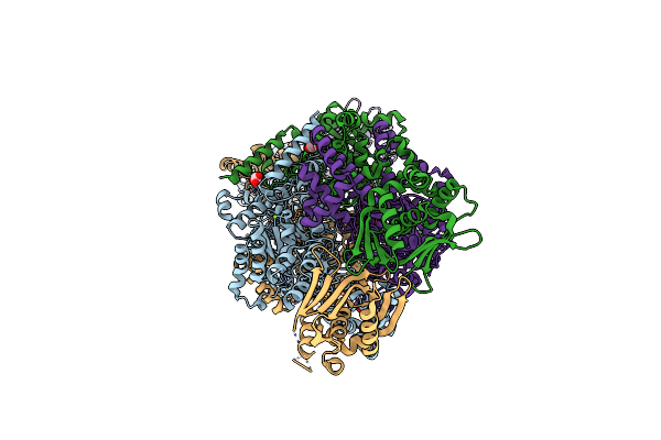
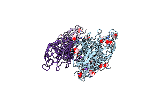
Crystal Structure Of M. Tuberculosis Glutamine-Dependent Nad+ Synthetase Complexed With Sulfonamide Derivative 1, Pyrophosphate, And Glutamine
Organism: Mycobacterium tuberculosis cdc1551
Method: X-RAY DIFFRACTION Resolution:3.14 Å Release Date: 2020-01-08 Classification: LIGASE Ligands: SFH, CL, POP, GLN, GOL |
 |
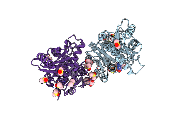
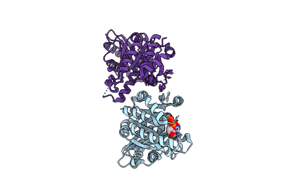
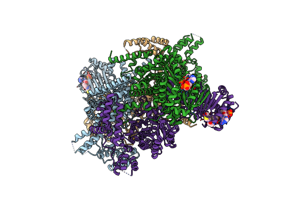
Structural Insights Into Mycobacterium Tuberculosis Clpp1P2 Inhibition By Cediranib: Implications For Developing Antimicrobial Agents Targeting Clp Protease
Organism: Mycobacterium tuberculosis cdc1551
Method: X-RAY DIFFRACTION Resolution:2.69 Å Release Date: 2020-01-01 Classification: HYDROLASE/HYDROLASE INHIBITOR Ligands: S0R, LEU, AV3 |
 |
Organism: Mycobacterium tuberculosis (strain cdc 1551 / oshkosh)
Method: X-RAY DIFFRACTION Resolution:1.80 Å Release Date: 2019-08-14 Classification: LYASE Ligands: GOL, MG |
 |
Crystal Structure Of Mycobacterium Tuberculosis Icl2 In Complex With Acetyl-Coa, Form I
Organism: Mycobacterium tuberculosis (strain cdc 1551 / oshkosh)
Method: X-RAY DIFFRACTION Resolution:2.67 Å Release Date: 2019-08-14 Classification: LYASE Ligands: ACO |
 |
Crystal Structure Of Mycobacterium Tuberculosis Icl2 In Complex With Acetyl-Coa
Organism: Mycobacterium tuberculosis (strain cdc 1551 / oshkosh)
Method: X-RAY DIFFRACTION Resolution:2.36 Å Release Date: 2019-08-14 Classification: LYASE Ligands: ACO, MG |
 |
Organism: Mycobacterium tuberculosis (strain cdc 1551 / oshkosh)
Method: X-RAY DIFFRACTION Resolution:1.51 Å Release Date: 2019-08-14 Classification: ANTIMICROBIAL PROTEIN Ligands: NO3, TRS, EDO, NA |
 |
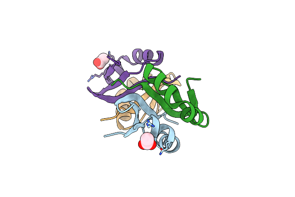
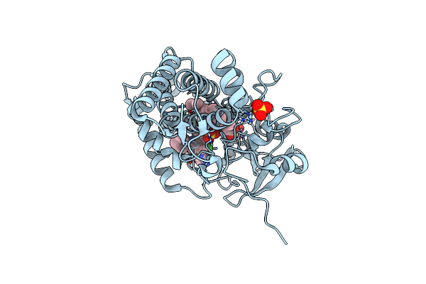
Crystal Structure Of Ldtmt2 From Mycobacterium Tuberculosis Bound To Ebselen
Organism: Mycobacterium tuberculosis cdc1551
Method: X-RAY DIFFRACTION Resolution:1.64 Å Release Date: 2019-08-14 Classification: ANTIMICROBIAL PROTEIN Ligands: NH4, GOL, 9JT, EDO |
 |
Crystal Structure Of Mycobacterium Tuberculosis Cytochrome P450 Cyp121A1 In Complex With Triazole Pyrazole Inhibitor 10J
Organism: Mycobacterium tuberculosis (strain cdc 1551 / oshkosh)
Method: X-RAY DIFFRACTION Resolution:1.50 Å Release Date: 2019-08-07 Classification: OXIDOREDUCTASE Ligands: HEM, SO4, EW2 |
 |
Crystal Structure Of Mycobacterium Tuberculosis Cytochrome P450 Cyp121A1 In Complex With Triazole Pyrazole Inhibitor 14A
Organism: Mycobacterium tuberculosis (strain cdc 1551 / oshkosh)
Method: X-RAY DIFFRACTION Resolution:1.60 Å Release Date: 2019-08-07 Classification: OXIDOREDUCTASE Ligands: HEM, EW5, SO4 |
 |
Inhibitor Bound Crystal Structure Of N-Terminal Domain Of Facl13 From Mycobacterium Tuberculosis
Organism: Mycobacterium tuberculosis cdc1551
Method: X-RAY DIFFRACTION Resolution:2.37 Å Release Date: 2019-04-24 Classification: LIGASE Ligands: JSA |