
Search Count: 64
 |
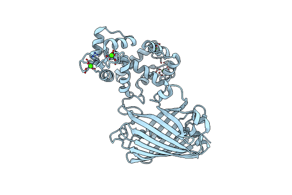
The Full-Length Human Sweet Taste Receptor Tas1R2 And Tas1R3 In The Apo State
Organism: Homo sapiens, Discosoma sp. (sea anemone), Branchiostoma lanceolatum
Method: ELECTRON MICROSCOPY Resolution:3.41 Å Release Date: 2025-07-16 Classification: SIGNALING PROTEIN |
 |
The Vft Domains Of Human Sweet Taste Receptor Tas1R2 And Tas1R3 In The Apo State
Organism: Homo sapiens, Discosoma sp. (sea anemone), Branchiostoma lanceolatum
Method: ELECTRON MICROSCOPY Resolution:3.18 Å Release Date: 2025-07-16 Classification: SIGNALING PROTEIN |
 |
The Transmembrane Domains Of Human Sweet Taste Receptor Tas1R2 And Tas1R3 In The Apo State
Organism: Homo sapiens, Discosoma sp. (sea anemone), Branchiostoma lanceolatum
Method: ELECTRON MICROSCOPY Resolution:3.77 Å Release Date: 2025-07-16 Classification: SIGNALING PROTEIN |
 |
The Full-Length Human Sweet Taste Receptor Tas1R2 And Tas1R3 In The Sucralose-Bound State
Organism: Homo sapiens, Discosoma sp., Branchiostoma lanceolatum
Method: ELECTRON MICROSCOPY Resolution:3.59 Å Release Date: 2025-07-16 Classification: SIGNALING PROTEIN |
 |
The Vft Domains Of Human Sweet Taste Receptor Tas1R2 And Tas1R3 In The Sucralose-Bound State
Organism: Homo sapiens, Discosoma sp., Branchiostoma lanceolatum
Method: ELECTRON MICROSCOPY Resolution:3.33 Å Release Date: 2025-07-16 Classification: SIGNALING PROTEIN |
 |
Organism: Branchiostoma lanceolatum
Method: X-RAY DIFFRACTION Resolution:2.32 Å Release Date: 2025-06-04 Classification: FLUORESCENT PROTEIN Ligands: SO4 |
 |
Organism: Escherichia coli o157:h7, Branchiostoma lanceolatum
Method: X-RAY DIFFRACTION Resolution:2.65 Å Release Date: 2024-12-25 Classification: FLUORESCENT PROTEIN Ligands: ILE, GOL, CL |
 |
Organism: Enterobacteria phage m13
Method: ELECTRON MICROSCOPY Release Date: 2024-01-03 Classification: VIRAL PROTEIN |
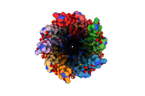 |
Organism: Enterobacteria phage m13
Method: ELECTRON MICROSCOPY Release Date: 2024-01-03 Classification: VIRAL PROTEIN |
 |
Organism: Branchiostoma lanceolatum
Method: X-RAY DIFFRACTION Resolution:1.28 Å Release Date: 2023-11-15 Classification: BIOSYNTHETIC PROTEIN Ligands: CA, SO4 |
 |
Organism: Inovirus m13
Method: ELECTRON MICROSCOPY Release Date: 2023-09-13 Classification: VIRAL PROTEIN |
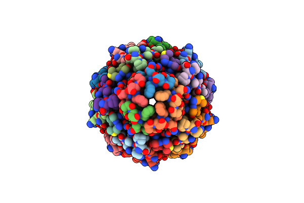 |
Organism: Inovirus m13
Method: ELECTRON MICROSCOPY Release Date: 2023-09-13 Classification: VIRAL PROTEIN |
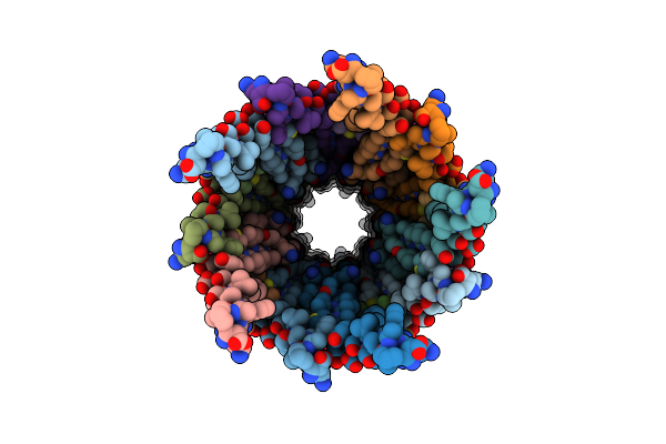 |
Organism: Inovirus m13
Method: ELECTRON MICROSCOPY Release Date: 2023-08-16 Classification: VIRAL PROTEIN |
 |
Organism: Enterobacteria phage m13
Method: ELECTRON MICROSCOPY Release Date: 2023-08-16 Classification: VIRAL PROTEIN |
 |
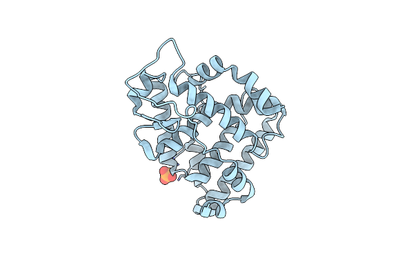
Genetically Encoded Green Ratiometric Calcium Indicator Fncamp In Calcium-Bound State
Organism: Branchiostoma lanceolatum, Aspergillus niger
Method: X-RAY DIFFRACTION Resolution:2.42 Å Release Date: 2023-05-24 Classification: FLUORESCENT PROTEIN Ligands: CA |
 |
The Crystallographic Structure Of The Ligand Binding Domain Of The Nr7 Nuclear Receptor From The Amphioxus Branchiostoma Lanceolatum
Organism: Branchiostoma lanceolatum
Method: X-RAY DIFFRACTION Resolution:2.00 Å Release Date: 2022-09-14 Classification: TRANSCRIPTION Ligands: CL, PO4 |
 |
Organism: Branchiostoma lanceolatum
Method: X-RAY DIFFRACTION Resolution:1.51 Å Release Date: 2022-05-04 Classification: FLUORESCENT PROTEIN |
 |
Organism: Branchiostoma lanceolatum
Method: X-RAY DIFFRACTION Resolution:1.95 Å Release Date: 2022-05-04 Classification: FLUORESCENT PROTEIN Ligands: SO4 |
 |
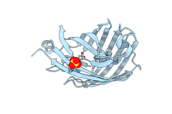
Structure Of The T207D Single-Point Mutant Of The Fluorescent Protein Neoncyan1 At Ph 6.5
Organism: Branchiostoma lanceolatum
Method: X-RAY DIFFRACTION Resolution:1.60 Å Release Date: 2022-05-04 Classification: FLUORESCENT PROTEIN |
 |
Structure Of The Yellow-Green Fluorescent Protein Mneongreen From Branchiostoma Lanceolatum At The Acidic Ph 4.5
Organism: Branchiostoma lanceolatum
Method: X-RAY DIFFRACTION Resolution:1.70 Å Release Date: 2016-12-21 Classification: FLUORESCENT PROTEIN Ligands: CL, FLC |