Planned Maintenance: Some services may turn out to be unavailable from 15th January, 2026 to 16th January, 2026. We apologize for the inconvenience!

Planned Maintenance: Some services may turn out to be unavailable from 15th January, 2026 to 16th January, 2026. We apologize for the inconvenience!
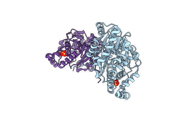 |
Organism: Mannheimia haemolytica
Method: X-RAY DIFFRACTION Resolution:2.20 Å Release Date: 2017-08-02 Classification: MEMBRANE PROTEIN Ligands: SO4 |
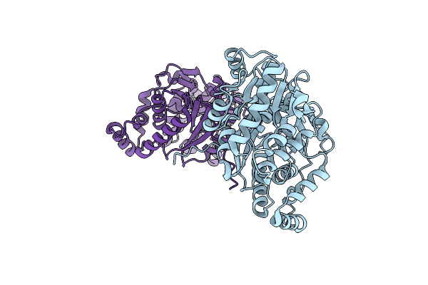 |
Organism: Mannheimia haemolytica
Method: X-RAY DIFFRACTION Resolution:2.75 Å Release Date: 2017-08-02 Classification: MEMBRANE PROTEIN Ligands: BR |
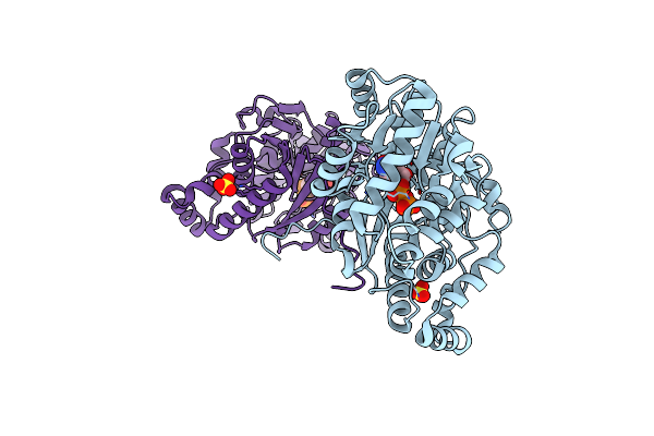 |
Organism: Mannheimia haemolytica
Method: X-RAY DIFFRACTION Resolution:3.00 Å Release Date: 2017-08-02 Classification: MEMBRANE PROTEIN Ligands: SO4, CDP |
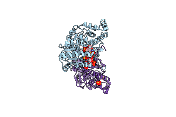 |
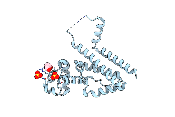
Structure Of A Bacterial Polysialyltransferase In Complex With Fondaparinux
Organism: Mannheimia haemolytica
Method: X-RAY DIFFRACTION Resolution:3.10 Å Release Date: 2017-08-02 Classification: MEMBRANE PROTEIN Ligands: SO4 |
 |
Organism: Mannheimia haemolytica
Method: X-RAY DIFFRACTION Resolution:2.20 Å Release Date: 2016-01-20 Classification: TRANSCRIPTION Ligands: SO4, CL, GOL |
 |
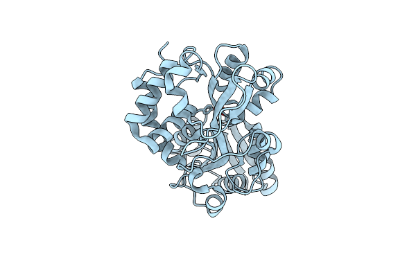
Crystal Structure Of Mannheimia Haemolytica Ferric Iron-Binding Protein A In A Closed Conformation
Organism: Mannheimia haemolytica
Method: X-RAY DIFFRACTION Resolution:1.35 Å Release Date: 2004-06-08 Classification: METAL BINDING PROTEIN Ligands: FE, CO3, EDO |
 |
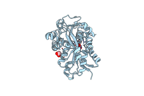
Crystal Structure Of Mannheimia Haemolytica Ferric Iron-Binding Protein A In An Open Conformation
Organism: Mannheimia haemolytica
Method: X-RAY DIFFRACTION Resolution:1.45 Å Release Date: 2004-06-08 Classification: METAL BINDING PROTEIN Ligands: FE |
 |
Crystal Structure Of Pasteurella Haemolytica Apo Ferric Ion-Binding Protein A
Organism: Mannheimia haemolytica
Method: X-RAY DIFFRACTION Resolution:1.20 Å Release Date: 2003-11-11 Classification: METAL BINDING PROTEIN Ligands: EDO, FMT |
 |
 |
Organism: Cosmopteriginae sp. BIOUG02374-G09
Method: Alphafold Release Date: Classification: NA Ligands: NA |
 |
NA
|
 |
NA
|
 |
NA
|
 |
NA
|
 |
NA
|
 |
NA
|
 |
NA
|
 |
NA
|
 |
NA
|
 |
NA
|