
Search Count: 2,462
 |
Organism: Caloramator australicus rc3, Unidentified
Method: X-RAY DIFFRACTION Release Date: 2025-12-10 Classification: METAL BINDING PROTEIN Ligands: ZN, NA, EPE, MG |
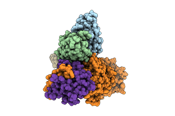 |
Cryo-Em Structure Of Kshv Glycoprotein Ghgl In Complex With Mlkh3 And Mlkh10 Fabs
Organism: Mus musculus, Human gammaherpesvirus 8
Method: ELECTRON MICROSCOPY Release Date: 2025-12-10 Classification: IMMUNE SYSTEM |
 |
Organism: Homo sapiens, Human gammaherpesvirus 8, Mus musculus
Method: ELECTRON MICROSCOPY Release Date: 2025-09-03 Classification: VIRAL PROTEIN/SIGNALING PROTEIN |
 |
Organism: Human gammaherpesvirus 8
Method: ELECTRON MICROSCOPY Release Date: 2025-08-27 Classification: VIRAL PROTEIN |
 |
Cryo-Em Structure Of The Light-Driven Proton Pump Pspr In Detergent Micelle
Organism: Candidatus pseudothioglobus sp.
Method: ELECTRON MICROSCOPY Release Date: 2025-07-23 Classification: MEMBRANE PROTEIN Ligands: LFA, RET, LMT |
 |
Cryo-Em Structure Of The Double Mutant H84V/E120G Of The Flotillin-Associated Rhodopsin Psfar In Detergent Micelle
Organism: Candidatus pseudothioglobus sp.
Method: ELECTRON MICROSCOPY Release Date: 2025-07-23 Classification: MEMBRANE PROTEIN Ligands: LFA, RET |
 |
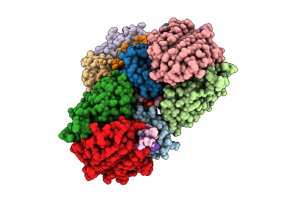
Structure Of R2 Retrotransposon Protein From Taeniopygia Guttata Initiating Target-Primed Reverse Transcription
Organism: Taeniopygia guttata, Synthetic construct
Method: ELECTRON MICROSCOPY Release Date: 2025-06-18 Classification: RNA BINDING PROTEIN/RNA/DNA Ligands: ZN, MG, TTP |
 |
Organism: Rhodobacter capsulatus sb 1003
Method: X-RAY DIFFRACTION Release Date: 2025-06-04 Classification: ELECTRON TRANSPORT Ligands: FES |
 |
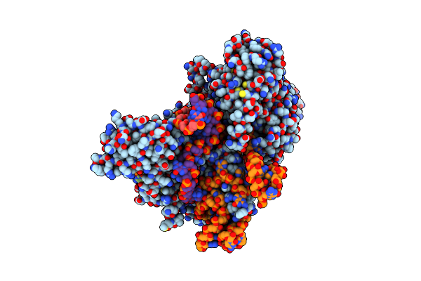
Taeniopygia Guttata R2 Retrotransposon (R2Tg) Initiating Target-Primed Reverse Transcription
Organism: Taeniopygia guttata
Method: ELECTRON MICROSCOPY Release Date: 2025-05-21 Classification: RNA BINDING PROTEIN/RNA/DNA Ligands: MG, TTP, ZN |
 |
Organism: Human gammaherpesvirus 8, Homo sapiens
Method: ELECTRON MICROSCOPY Release Date: 2025-03-05 Classification: VIRAL PROTEIN/IMMUNE SYSTEM |
 |
Organism: Human gammaherpesvirus 8
Method: ELECTRON MICROSCOPY Release Date: 2025-01-08 Classification: VIRAL PROTEIN Ligands: NAG |
 |
Crystal Structure Of The Catalytic Domain Of An Aa9 Lytic Polysaccharide Monooxygenase From Thermothelomyces Thermophilus (Ttlpmo9F)
Organism: Thermothelomyces thermophilus atcc 42464
Method: X-RAY DIFFRACTION Resolution:2.33 Å Release Date: 2024-12-25 Classification: METAL BINDING PROTEIN Ligands: PEG, PG4, EDO, FMT, CU |
 |
Cryo-Em Structure Of Kaposi'S Sarcoma-Associated Herpesvirus-G Protein-Coupled Receptor (Kshv-Gpcr)In Complex With Cxc Chemokine Cxcl1
Organism: Human gammaherpesvirus 8, Homo sapiens
Method: ELECTRON MICROSCOPY Release Date: 2024-07-24 Classification: VIRAL PROTEIN |
 |
Cryo-Em Structure Of An Active Kaposi'S Sarcoma-Associated Herpesvirus-G Protein-Coupled Receptor (Kshv-Gpcr) In Complex With Gi Protein
Organism: Homo sapiens, Human gammaherpesvirus 8
Method: ELECTRON MICROSCOPY Release Date: 2024-07-24 Classification: VIRAL PROTEIN |
 |
Organism: Neisseria meningitidis, Haemophilus parainfluenzae
Method: X-RAY DIFFRACTION Resolution:2.70 Å Release Date: 2024-01-17 Classification: ANTIMICROBIAL PROTEIN Ligands: HOH |
 |
Double Mutant A(L37)C/S(L99)C Structure Of Photosynthetic Reaction Center From Cereibacter Sphaeroides Strain Rv
Organism: Cereibacter sphaeroides 2.4.1
Method: X-RAY DIFFRACTION Resolution:2.60 Å Release Date: 2023-11-22 Classification: PHOTOSYNTHESIS Ligands: LDA, UNL, PO4, EDO, K, BCL, BPH, U10, DIO, HTO, CDL, FE, SPN |
 |
Double Mutant A(L53)C/I(L64)C Structure Of Photosynthetic Reaction Center From Cereibacter Sphaeroides Strain Rv
Organism: Cereibacter sphaeroides 2.4.1
Method: X-RAY DIFFRACTION Resolution:2.86 Å Release Date: 2023-11-22 Classification: PHOTOSYNTHESIS Ligands: LDA, UNL, PO4, EDO, K, BCL, BPH, U10, DIO, HTO, CDL, FE, SPN |
 |
Double Mutant V(M84)C/A(L278)C Structure Of Photosynthetic Reaction Center From Cereibacter Sphaeroides Strain Rv
Organism: Cereibacter sphaeroides 2.4.1
Method: X-RAY DIFFRACTION Resolution:2.60 Å Release Date: 2023-11-22 Classification: PHOTOSYNTHESIS Ligands: LDA, UNL, EDO, BCL, BPH, HTO, CDL, FE, U10, SPN |
 |
Double Mutant A(L172)C/L(L246)C Structure Of Photosynthetic Reaction Center From Cereibacter Sphaeroides Strain Rv
Organism: Cereibacter sphaeroides 2.4.1
Method: X-RAY DIFFRACTION Resolution:2.45 Å Release Date: 2023-11-22 Classification: PHOTOSYNTHESIS Ligands: LDA, UNL, EDO, OLC, BCL, BPH, HTO, CDL, FE, U10, SPN |
 |
Double Mutant G(M19)C/T(L214)C Structure Of Photosynthetic Reaction Center From Cereibacter Sphaeroides Strain Rv
Organism: Cereibacter sphaeroides 2.4.1
Method: X-RAY DIFFRACTION Resolution:2.75 Å Release Date: 2023-11-22 Classification: PHOTOSYNTHESIS Ligands: NKP, LDA, UNL, OLC, BCL, BPH, DIO, EDO, FE, U10, SPN, PO4 |