
Search Count: 5
 |
Organism: Paenibacillus alvei
Method: X-RAY DIFFRACTION Resolution:2.25 Å Release Date: 2018-08-15 Classification: SUGAR BINDING PROTEIN Ligands: 6LA |
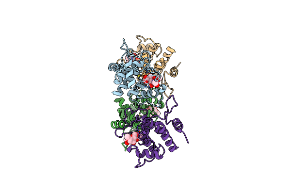 |
Crystal Structure Of Spaa-Slh In Complex With 4,6-Pyr-Beta-D-Mannacome (P1)
Organism: Paenibacillus alvei
Method: X-RAY DIFFRACTION Resolution:2.00 Å Release Date: 2018-08-15 Classification: SUGAR BINDING PROTEIN Ligands: 6LA |
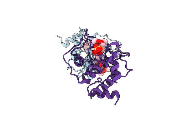 |
Crystal Structure Of Spaa-Slh In Complex With 4,6-Pyr-Beta-D-Mannacome (C2)
Organism: Paenibacillus alvei
Method: X-RAY DIFFRACTION Resolution:2.16 Å Release Date: 2018-08-15 Classification: SUGAR BINDING PROTEIN Ligands: 6LA, SO4 |
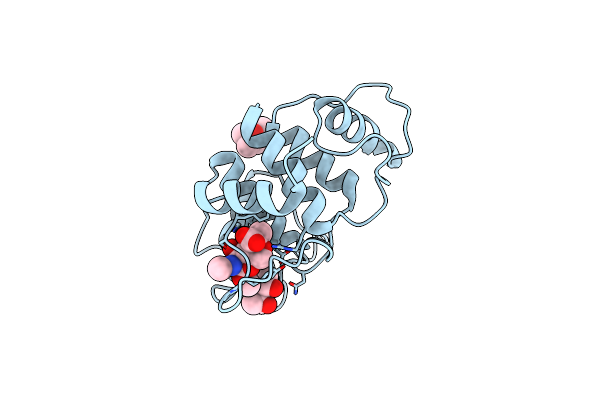 |
Crystal Structure Of Spaa-Slh/G109A In Complex With 4,6-Pyr-Beta-D-Mannacome
Organism: Paenibacillus alvei
Method: X-RAY DIFFRACTION Resolution:1.53 Å Release Date: 2018-08-15 Classification: SUGAR BINDING PROTEIN Ligands: 6LA, MPD |
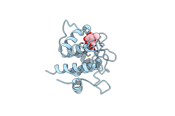 |
Crystal Structure Of Spaa-Slh/G46A/G109A In Complex With 4,6-Pyr-Beta-D-Mannacome
Organism: Paenibacillus alvei
Method: X-RAY DIFFRACTION Resolution:1.24 Å Release Date: 2018-08-15 Classification: SUGAR BINDING PROTEIN Ligands: 6LA |