
Search Count: 9
 |
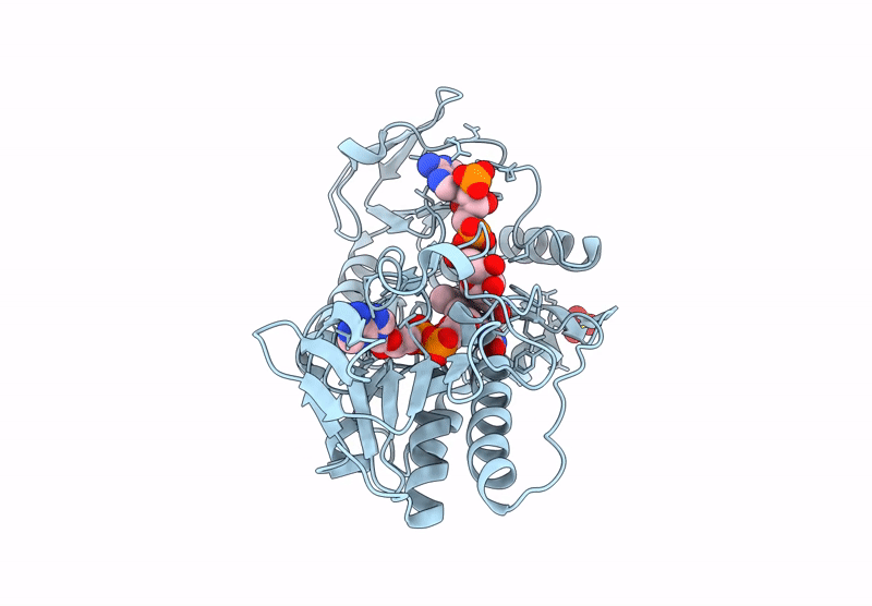
Organism: Homo sapiens
Method: X-RAY DIFFRACTION Resolution:2.00 Å Release Date: 2025-01-08 Classification: OXIDOREDUCTASE Ligands: SO4, 6FA, NAD |
 |
Organism: Homo sapiens
Method: X-RAY DIFFRACTION Resolution:1.79 Å Release Date: 2025-01-08 Classification: OXIDOREDUCTASE Ligands: 6FA, NDP, SO4 |
 |
Organism: Homo sapiens
Method: X-RAY DIFFRACTION Resolution:2.48 Å Release Date: 2025-01-08 Classification: OXIDOREDUCTASE Ligands: 6FA, SO4 |
 |
Organism: Homo sapiens
Method: X-RAY DIFFRACTION Resolution:2.00 Å Release Date: 2024-05-08 Classification: OXIDOREDUCTASE Ligands: 6FA, NAD |
 |
Organism: Homo sapiens
Method: X-RAY DIFFRACTION Resolution:2.16 Å Release Date: 2024-03-13 Classification: OXIDOREDUCTASE Ligands: 6FA, NAP |
 |
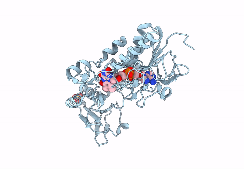
Crystal Structure Of Pseudomonas Aeruginosa D-Arginine Dehydrogenase Y249F Co-Crystallized In The Presence Of D-Arginine
Organism: Pseudomonas aeruginosa (strain atcc 15692 / dsm 22644 / cip 104116 / jcm 14847 / lmg 12228 / 1c / prs 101 / pao1)
Method: X-RAY DIFFRACTION Resolution:1.29 Å Release Date: 2021-12-22 Classification: FLAVOPROTEIN Ligands: 4R2, 6FA, GOL, PEG |
 |
Crystal Structure Of Pseudomonas Aeruginosa D-Arginine Dehydrogenase Y249F Variant With 6-Oh-Fad - Green Fraction
Organism: Pseudomonas aeruginosa (strain atcc 15692 / dsm 22644 / cip 104116 / jcm 14847 / lmg 12228 / 1c / prs 101 / pao1)
Method: X-RAY DIFFRACTION Resolution:1.55 Å Release Date: 2020-07-01 Classification: FLAVOPROTEIN Ligands: 6FA, PEG, GOL |
 |
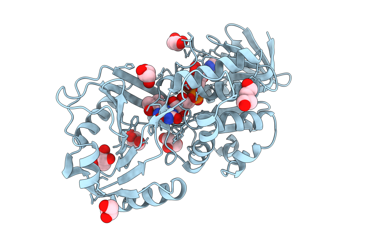
Cellobiose Dehydrogenase Flavoprotein Fragment In Complex With Cellobionolactam
Organism: Phanerochaete chrysosporium
Method: X-RAY DIFFRACTION Resolution:1.80 Å Release Date: 2003-01-14 Classification: OXIDOREDUCTASE Ligands: NAG, 6FA, ABL |
 |
Organism: Phanerochaete chrysosporium
Method: X-RAY DIFFRACTION Resolution:1.50 Å Release Date: 2002-11-13 Classification: OXIDOREDUCTASE Ligands: NAG, MAN, HG, 6FA, EMT, UNX |