Planned Maintenance: Some services may turn out to be unavailable from 15th January, 2026 to 16th January, 2026. We apologize for the inconvenience!

Planned Maintenance: Some services may turn out to be unavailable from 15th January, 2026 to 16th January, 2026. We apologize for the inconvenience!
 |
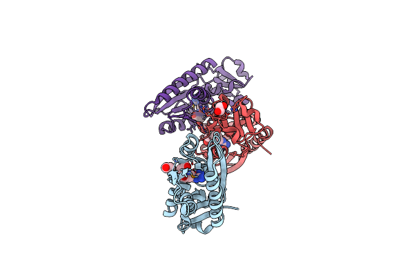
Structure Of The Periplasmic Binding Protein (Pbp) Occj From A. Tumefaciens B6 In Complex With Octopine.
Organism: Agrobacterium tumefaciens str. b6
Method: X-RAY DIFFRACTION Resolution:1.99 Å Release Date: 2017-12-20 Classification: Octopine-binding protein Ligands: EDO, NA, CL, GOL, 6DB, ACT |
 |
Structure Of The Periplasmic Binding Protein (Pbp) Noct-G97S Mutant From A. Tumefaciens C58 In Complex With Octopine.
Organism: Agrobacterium fabrum str. c58
Method: X-RAY DIFFRACTION Resolution:2.35 Å Release Date: 2017-12-20 Classification: PROTEIN BINDING Ligands: 6DB, EDO, SO4, CL, PEG |
 |
Crystal Structure Of The Ligand Binding Domain Of Lysr-Type Transcriptional Regulator, Occr From Agrobacterium Tumefaciens In The Complex With Octopine
Organism: Agrobacterium tumefaciens (strain ach5)
Method: X-RAY DIFFRACTION Resolution:2.28 Å Release Date: 2017-06-21 Classification: TRANSCRIPTION Ligands: 6DB, EDO, CL, ACY |
 |
Structure Of The Periplasmic Binding Protein M117N-Noct From A. Tumefaciens In Complex With Octopine
Organism: Agrobacterium fabrum (strain c58 / atcc 33970)
Method: X-RAY DIFFRACTION Resolution:2.35 Å Release Date: 2016-11-30 Classification: MEMBRANE PROTEIN Ligands: PEG, EDO, 1PE, 6DB, TOE |
 |
Structure Of The Periplasmic Binding Protein Noct From A.Tumefaciens In Complex With Octopine
Organism: Agrobacterium fabrum
Method: X-RAY DIFFRACTION Resolution:1.85 Å Release Date: 2016-11-30 Classification: MEMBRANE PROTEIN Ligands: 6DB, EDO, PEG |