
Search Count: 3,395
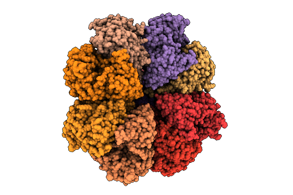 |
Organism: Streptococcus thermophilus, Streptococcus phage vb_sths-va214
Method: ELECTRON MICROSCOPY Release Date: 2025-11-12 Classification: RNA BINDING PROTEIN |
 |
Crystal Structure Of The Semet-Derived C-Terminal Of Viral Responsive Protein 15 (Pmvrp15) From Black Tiger Shrimp Penaeus Monodon
Organism: Penaeus monodon
Method: X-RAY DIFFRACTION Release Date: 2025-10-22 Classification: UNKNOWN FUNCTION Ligands: SO4 |
 |
Crystal Structure Of The C-Terminal Of Viral Responsive Protein 15 (Pmvrp15) From Black Tiger Shrimp Penaeus Monodon
Organism: Penaeus monodon
Method: X-RAY DIFFRACTION Release Date: 2025-10-22 Classification: UNKNOWN FUNCTION |
 |
Organism: Strongylocentrotus purpuratus
Method: ELECTRON MICROSCOPY Release Date: 2025-07-09 Classification: MEMBRANE PROTEIN |
 |
Organism: Strongylocentrotus purpuratus
Method: ELECTRON MICROSCOPY Release Date: 2025-03-26 Classification: MEMBRANE PROTEIN Ligands: POV, AJP |
 |
Organism: Strongylocentrotus purpuratus
Method: ELECTRON MICROSCOPY Release Date: 2025-03-26 Classification: MEMBRANE PROTEIN Ligands: POV, AJP |
 |
Organism: Strongylocentrotus purpuratus
Method: ELECTRON MICROSCOPY Release Date: 2025-03-26 Classification: MEMBRANE PROTEIN Ligands: POV, AJP |
 |
Organism: Strongylocentrotus purpuratus
Method: ELECTRON MICROSCOPY Release Date: 2025-03-26 Classification: MEMBRANE PROTEIN Ligands: POV, AJP |
 |
Organism: Strongylocentrotus purpuratus
Method: ELECTRON MICROSCOPY Release Date: 2025-03-26 Classification: MEMBRANE PROTEIN Ligands: POV, AJP |
 |
Organism: Strongylocentrotus purpuratus
Method: ELECTRON MICROSCOPY Release Date: 2025-03-26 Classification: MEMBRANE PROTEIN Ligands: POV, AJP, CMP |
 |
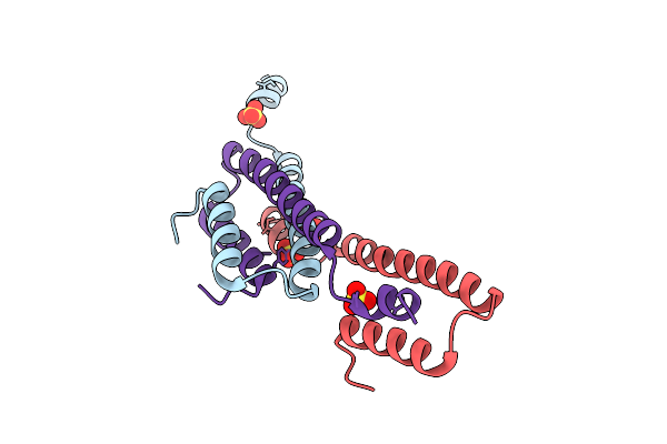
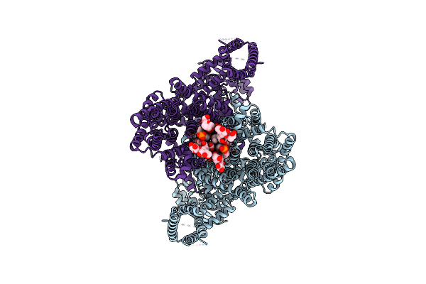
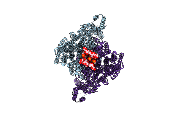
Wt Sea Urchin Slc9C1 With 5Mm Camp At Ph 6 In Na+ - Grip And Twist (Gnt) Conformation
Organism: Strongylocentrotus purpuratus
Method: ELECTRON MICROSCOPY Release Date: 2025-03-26 Classification: MEMBRANE PROTEIN Ligands: POV, AJP, CMP |
 |
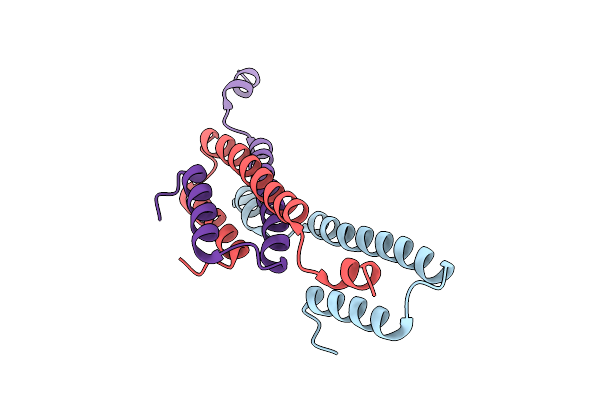
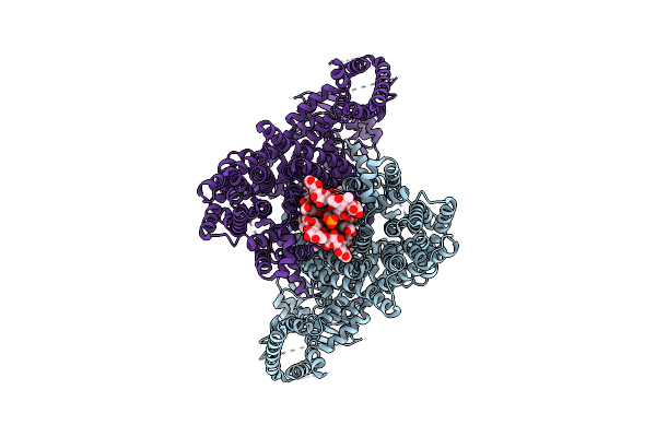
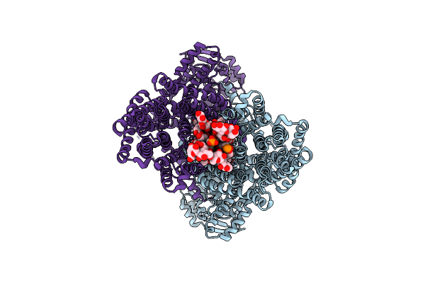
Wt Sea Urchin Slc9C1 With 5Mm Camp At Ph 6 In Na+ - Grip And Twist Like (Gntl) Conformation
Organism: Strongylocentrotus purpuratus
Method: ELECTRON MICROSCOPY Release Date: 2025-03-26 Classification: MEMBRANE PROTEIN Ligands: POV, AJP, CMP |
 |
Organism: Strongylocentrotus purpuratus
Method: ELECTRON MICROSCOPY Release Date: 2025-03-26 Classification: MEMBRANE PROTEIN Ligands: POV, AJP |
 |
Organism: Strongylocentrotus purpuratus
Method: ELECTRON MICROSCOPY Release Date: 2025-03-26 Classification: MEMBRANE PROTEIN Ligands: POV, AJP |
 |
Crystal Structure Of Gh65 Alpha-1,2-Glucosidase From Flavobacterium Johnsoniae In Complex With 1-Deoxynojirimycin
Organism: Flavobacterium johnsoniae uw101
Method: X-RAY DIFFRACTION Resolution:1.90 Å Release Date: 2025-03-19 Classification: HYDROLASE Ligands: NOJ, EDO |
 |
Crystal Structure Of Gh65 Alpha-1,2-Glucosidase From Flavobacterium Johnsoniae In Complex With Castanospermine
Organism: Flavobacterium johnsoniae uw101
Method: X-RAY DIFFRACTION Resolution:1.60 Å Release Date: 2025-03-19 Classification: HYDROLASE Ligands: CTS, EDO |
 |
Crystal Structure Of Sm0281, An Alpha/Beta Hydrolase From Sinorhizobium Meliloti
Organism: Rhizobium meliloti (strain 1021)
Method: X-RAY DIFFRACTION Resolution:2.46 Å Release Date: 2025-02-12 Classification: HYDROLASE Ligands: GOL, PE3, CL |
 |
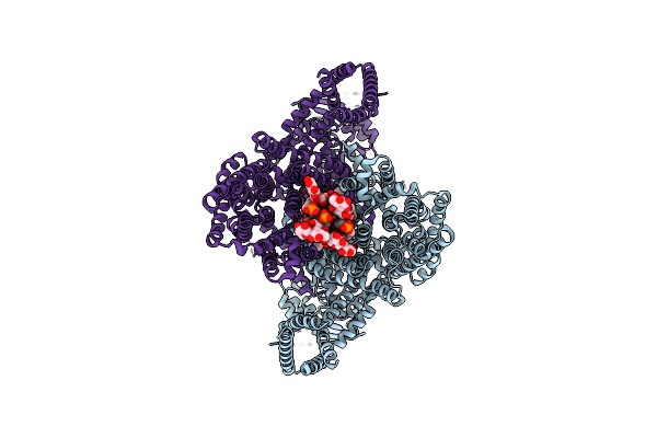
Organism: Strongylocentrotus purpuratus
Method: ELECTRON MICROSCOPY Release Date: 2025-01-15 Classification: TRANSPORT PROTEIN |
 |
Organism: Strongylocentrotus purpuratus
Method: ELECTRON MICROSCOPY Release Date: 2025-01-15 Classification: TRANSPORT PROTEIN |
 |
Organism: Streptomyces sp. atcc 700974
Method: X-RAY DIFFRACTION Resolution:2.50 Å Release Date: 2024-10-09 Classification: OXIDOREDUCTASE Ligands: SF4, SO4 |