
Search Count: 3,118
 |
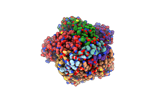
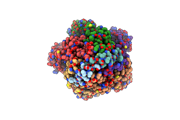
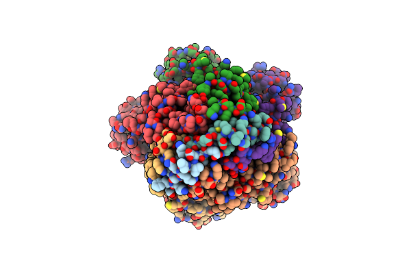
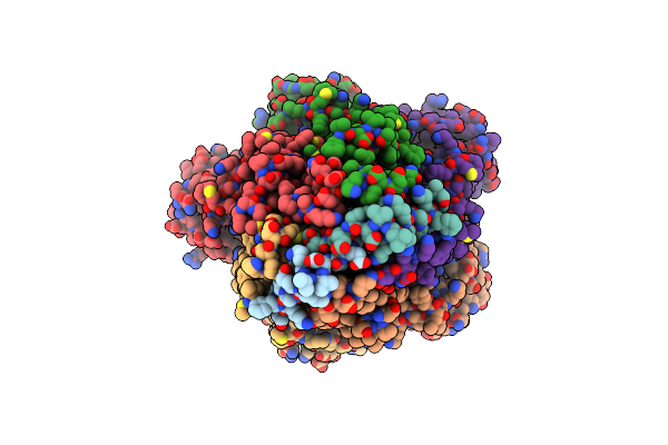
Mutant H286T Crystal Structure Of Two-Domain Bacterial Laccase From The Actinobacterium Streptomyces Carpinensis Vkm Ac-1300
Organism: Streptomyces carpinensis
Method: X-RAY DIFFRACTION Resolution:2.05 Å Release Date: 2025-08-06 Classification: OXIDOREDUCTASE Ligands: CU, OXY, GOL |
 |
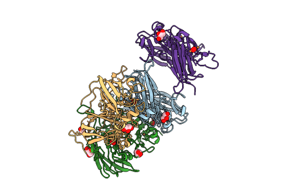
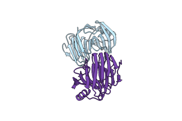
Molecular Basis Of Pathogenicity Of The Recently Emerged Fcov-23 Coronavirus. Complex Of Fapn With Fcov-23 Rbd
Organism: Felis catus, Feline coronavirus
Method: ELECTRON MICROSCOPY Release Date: 2025-07-09 Classification: VIRAL PROTEIN/HYDROLASE Ligands: NAG, ZN |
 |
Molecular Basis Of Pathogenicity Of The Recently Emerged Fcov-23 Coronavirus. Fcov-23 S Short
Organism: Feline coronavirus
Method: ELECTRON MICROSCOPY Release Date: 2025-07-09 Classification: VIRAL PROTEIN Ligands: NAG, PAM |
 |
Molecular Basis Of Pathogenicity Of The Recently Emerged Fcov-23 Coronavirus. Fcov-23 S Do In Proximal Conformation (Local Refinement)
Organism: Feline coronavirus
Method: ELECTRON MICROSCOPY Release Date: 2025-07-09 Classification: VIRAL PROTEIN Ligands: NAG |
 |
Molecular Basis Of Pathogenicity Of The Recently Emerged Fcov-23 Coronavirus. Fcov-23 S Long With Do In Swung-Out Conformation
Organism: Feline coronavirus
Method: ELECTRON MICROSCOPY Release Date: 2025-07-09 Classification: VIRAL PROTEIN Ligands: NAG, PAM |
 |
Molecular Basis Of Pathogenicity Of The Recently Emerged Fcov-23 Coronavirus. Fcov-23 S Long Domain 0 In Swung-Out Conformation (Local Refinement)
Organism: Feline coronavirus
Method: ELECTRON MICROSCOPY Release Date: 2025-07-09 Classification: VIRAL PROTEIN Ligands: NAG |
 |
Molecular Basis Of Pathogenicity Of The Recently Emerged Fcov-23 Coronavirus. Fcov-23 S Long With Do In Mixed Conformations (Global Refinement).
Organism: Feline coronavirus
Method: ELECTRON MICROSCOPY Release Date: 2025-07-09 Classification: VIRAL PROTEIN Ligands: NAG, PAM |
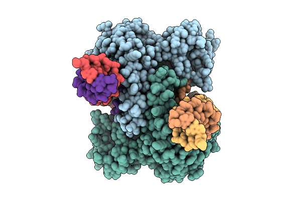 |
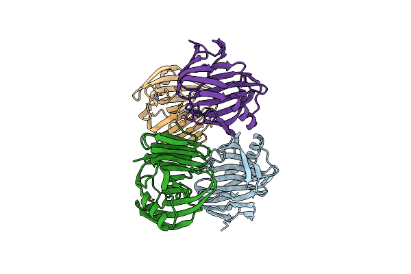
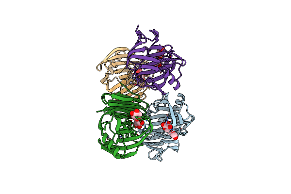
Structure Of Tigr-Tasr In Complex With Tigrna And Target Dna After Dna Cleavage
Organism: Thermoproteota archaeon, Escherichia coli
Method: ELECTRON MICROSCOPY Resolution:3.05 Å Release Date: 2025-03-05 Classification: DNA BINDING PROTEIN/RNA/DNA Ligands: MG |
 |
Organism: Escherichia coli, Vibrio alginolyticus
Method: ELECTRON MICROSCOPY Release Date: 2025-02-12 Classification: RIBOSOME |
 |
Organism: Escherichia coli, Vibrio alginolyticus
Method: ELECTRON MICROSCOPY Release Date: 2025-02-12 Classification: RIBOSOME |
 |
Crystal Structure Of Ferric Iron-Binding Protein (Fecb) From Vibrio Alginolyticus In Complex With Citrate
Organism: Vibrio alginolyticus
Method: X-RAY DIFFRACTION Resolution:2.31 Å Release Date: 2025-02-05 Classification: METAL BINDING PROTEIN Ligands: FLC |
 |
Crystal Structure Of Ferric Iron-Binding Protein (Fecb) From Vibrio Alginolyticus In Complex With Ferric Citrate
Organism: Vibrio alginolyticus
Method: X-RAY DIFFRACTION Resolution:2.51 Å Release Date: 2025-02-05 Classification: METAL BINDING PROTEIN Ligands: GOL, PEG, FLC, FE |
 |
Organism: Vibrio alginolyticus
Method: ELECTRON MICROSCOPY Release Date: 2025-01-01 Classification: MEMBRANE PROTEIN |
 |
Organism: Vibrio alginolyticus
Method: ELECTRON MICROSCOPY Release Date: 2025-01-01 Classification: MEMBRANE PROTEIN |
 |
Organism: Vibrio alginolyticus
Method: ELECTRON MICROSCOPY Release Date: 2025-01-01 Classification: MEMBRANE PROTEIN |
 |
Organism: Vibrio alginolyticus
Method: ELECTRON MICROSCOPY Release Date: 2025-01-01 Classification: MEMBRANE PROTEIN |
 |
Bacterial Flagellar Sodium-Driven Stator Poma5Pomb2 With 100 Mm Nacl And 0.1 Mm Phenamil
Organism: Vibrio alginolyticus
Method: ELECTRON MICROSCOPY Release Date: 2025-01-01 Classification: MEMBRANE PROTEIN |
 |
Organism: Aspergillus terreus (strain nih 2624 / fgsc a1156)
Method: X-RAY DIFFRACTION Resolution:2.30 Å Release Date: 2024-11-27 Classification: HYDROLASE |
 |
Organism: Aspergillus terreus (strain nih 2624 / fgsc a1156)
Method: X-RAY DIFFRACTION Resolution:1.75 Å Release Date: 2024-11-27 Classification: HYDROLASE Ligands: ZN |
 |
Organism: Aspergillus terreus nih2624
Method: X-RAY DIFFRACTION Resolution:1.72 Å Release Date: 2024-11-27 Classification: HYDROLASE Ligands: ZN, XYP |