Planned Maintenance: Some services may turn out to be unavailable from 15th January, 2026 to 16th January, 2026. We apologize for the inconvenience!

Planned Maintenance: Some services may turn out to be unavailable from 15th January, 2026 to 16th January, 2026. We apologize for the inconvenience!
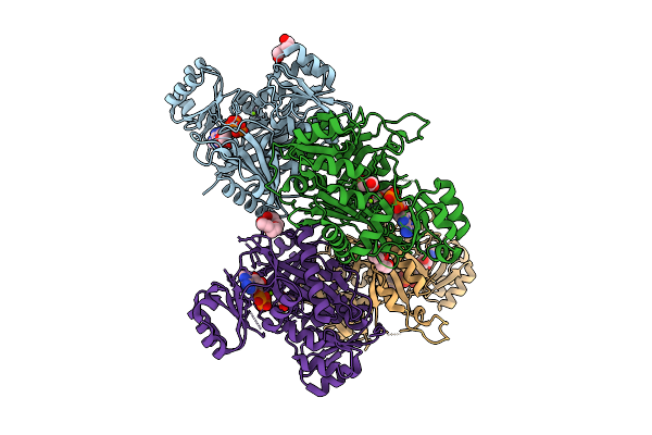 |
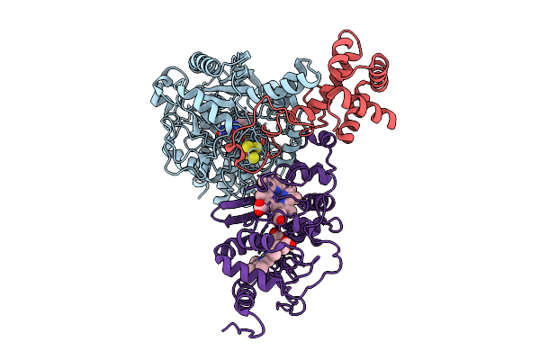
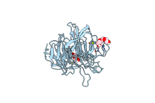
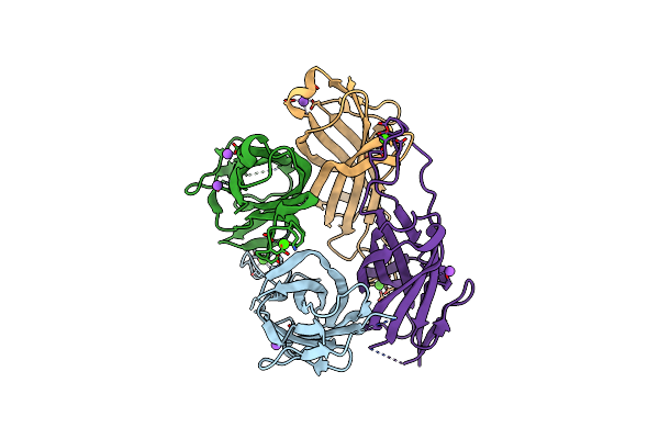
Crystal Structure Of Phosphoribosylaminoimidazole Carboxylase From Burkholderia Xenovorans (Atp Complex)
Organism: Paraburkholderia xenovorans lb400
Method: X-RAY DIFFRACTION Release Date: 2025-12-24 Classification: LYASE Ligands: ATP, PEG, MG, PGE, GOL, ADP, PG4 |
 |
Crystal Structure Of Phosphoribosylaminoimidazole Carboxylase From Burkholderia Xenovorans (Adp Complex)
Organism: Paraburkholderia xenovorans lb400
Method: X-RAY DIFFRACTION Release Date: 2025-12-24 Classification: LYASE Ligands: ADP, PEG, MG, PGE, GOL, PG4 |
 |
Crystal Structure Of Phosphoribosylaminoimidazole Carboxylase From Burkholderia Xenovorans (Amp Complex)
Organism: Paraburkholderia xenovorans lb400
Method: X-RAY DIFFRACTION Release Date: 2025-12-24 Classification: LYASE Ligands: AMP, PGE, MG, PEG, PG4, GOL |
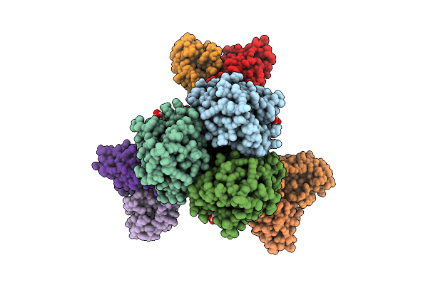 |
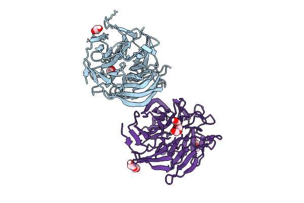
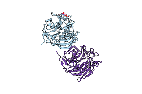
Organism: Homo sapiens, Influenza a virus (a/california/01/2009(h1n1))
Method: ELECTRON MICROSCOPY Release Date: 2025-11-12 Classification: IMMUNE SYSTEM/VIRAL PROTEIN Ligands: NAG |
 |
Organism: Homo sapiens, Influenza a virus (a/california/01/2009(h1n1))
Method: ELECTRON MICROSCOPY Release Date: 2025-11-12 Classification: IMMUNE SYSTEM/VIRAL PROTEIN Ligands: NAG |
 |
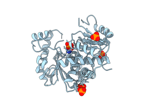
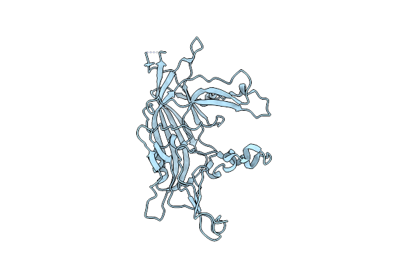
Crystal Structure Of Ornithine Carbamoyltransferase From Burkholderia Xenovorans
Organism: Paraburkholderia xenovorans lb400
Method: X-RAY DIFFRACTION Release Date: 2025-09-24 Classification: TRANSFERASE Ligands: CL |
 |
Crystal Structure Of Ornithine Carbamoyltransferase From Burkholderia Xenovorans In Complex With Phosphono Carbamate
Organism: Paraburkholderia xenovorans lb400
Method: X-RAY DIFFRACTION Release Date: 2025-09-24 Classification: TRANSFERASE Ligands: CP, PO4, CL |
 |
Cryo-Em Structure Of Fructose Dehydrogenase Variant From Gluconobacter Japonicus Truncating Heme 1C And C-Terminal Hydrophobic Regions
Organism: Gluconobacter japonicus
Method: ELECTRON MICROSCOPY Release Date: 2025-08-06 Classification: OXIDOREDUCTASE Ligands: FAD, F3S, HEC |
 |
Organism: Corynebacterium glutamicum atcc 13032
Method: X-RAY DIFFRACTION Release Date: 2025-07-30 Classification: CELL CYCLE |
 |
Crystal Structure Of Complement1.4 (Cent1.4), An Engineered Photoenzyme For Selective [2+2]-Cycloadditions
Organism: Loligo vulgaris
Method: X-RAY DIFFRACTION Release Date: 2025-07-16 Classification: BIOSYNTHETIC PROTEIN Ligands: EDO, PEG, PGE |
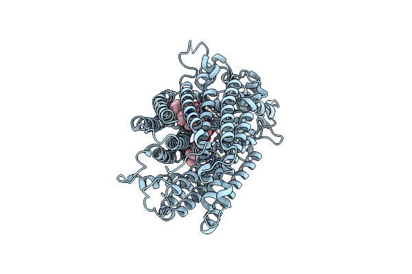 |
Cryoem Structure Of Monomeric Quinol Dependent Nitric Oxide Reductase From Neisseria Meningitidis
Organism: Neisseria meningitidis alpha14
Method: ELECTRON MICROSCOPY Release Date: 2025-05-14 Classification: OXIDOREDUCTASE Ligands: HEM, FE, CA |
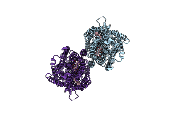 |
Cryoem Structure Of Dimeric Quinol Dependent Nitric Oxide Reductase From Neisseria Meningitidis
Organism: Neisseria meningitidis alpha14
Method: ELECTRON MICROSCOPY Release Date: 2025-05-14 Classification: OXIDOREDUCTASE Ligands: HEM, FE, CA |
 |
Organism: Loligo vulgaris
Method: X-RAY DIFFRACTION Release Date: 2025-05-14 Classification: BIOSYNTHETIC PROTEIN Ligands: EDO |
 |
Organism: Loligo vulgaris
Method: X-RAY DIFFRACTION Release Date: 2025-05-14 Classification: BIOSYNTHETIC PROTEIN Ligands: PEG, EDO |
 |
Organism: Loligo vulgaris
Method: X-RAY DIFFRACTION Release Date: 2025-05-14 Classification: BIOSYNTHETIC PROTEIN Ligands: PEG, PGE, MG |
 |
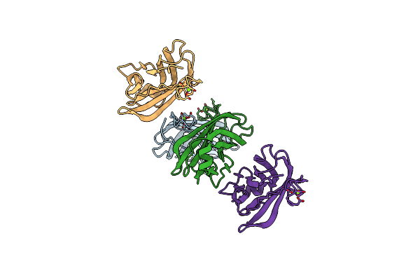
Capsid Structure Of The Syngnathus Scovelli Chapparvovirus Virus-Like Particle
Organism: Syngnathus scovelli chapparvovirus
Method: ELECTRON MICROSCOPY Release Date: 2025-03-05 Classification: VIRUS LIKE PARTICLE |
 |
Corynebacterium Glutamicum Pyruvate:Quinone Oxidoreductase (Pqo), C-Terminal Truncated Construct
Organism: Corynebacterium glutamicum atcc 13032
Method: X-RAY DIFFRACTION Resolution:1.89 Å Release Date: 2024-08-07 Classification: OXIDOREDUCTASE Ligands: FAD, TPP, MG |
 |
Intramolecular Ester Bond-Containing Repeat Domain 2 From Suipraoptans Intestinalis Adhesin
Organism: Suipraeoptans intestinalis
Method: X-RAY DIFFRACTION Resolution:1.60 Å Release Date: 2024-06-19 Classification: CELL ADHESION Ligands: MG |
 |
Intramolecular Ester Bond-Containing Repeat Domain 1 From Suipraoptans Intestinalis Adhesin
Organism: Suipraeoptans intestinalis
Method: X-RAY DIFFRACTION Resolution:1.49 Å Release Date: 2024-06-19 Classification: CELL ADHESION Ligands: CA, NA |
 |
Cryo-Em Structure Of Membrane-Bound Fructose Dehydrogenase From Gluconobacter Japonicus Variant-H1147A
Organism: Gluconobacter japonicus
Method: ELECTRON MICROSCOPY Release Date: 2024-05-22 Classification: OXIDOREDUCTASE Ligands: FAD, F3S, HEC, U10 |