
Search Count: 773
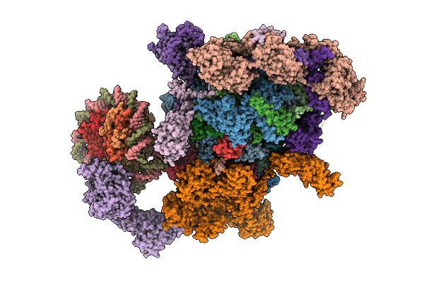 |
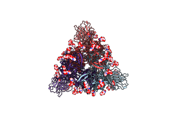
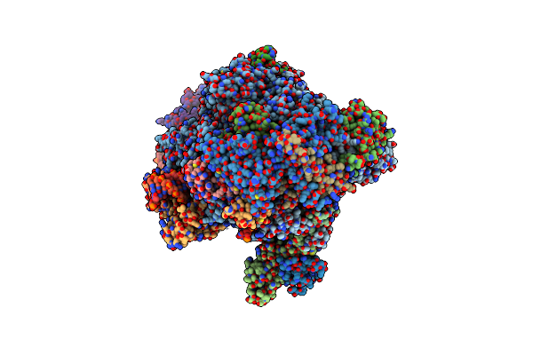
Co-Transcriptional Histone H3K36 Methylation Complex Containing Rna Polymerase Ii Elongation Complex, Set2, And The Upstream Nucleosome. (Temp115, Type B)
Organism: Komagataella phaffii gs115, Homo sapiens, Synthetic construct
Method: ELECTRON MICROSCOPY Release Date: 2025-12-24 Classification: TRANSCRIPTION Ligands: ZN, MG, SAH |
 |
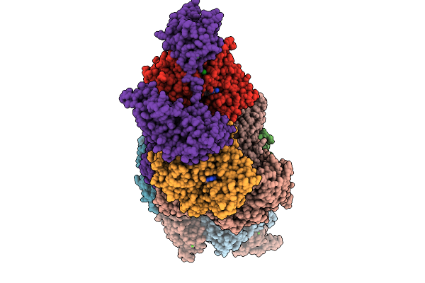
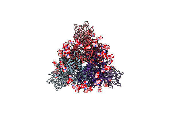
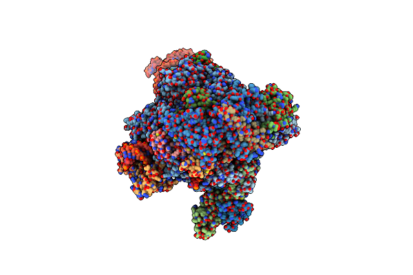
Co-Transcriptional Histone H3K36 Methylation Complex Containing Rna Polymerase Ii Elongation Complex, Set2, And The Upstream Nucleosome. (Temp115, Type A)
Organism: Komagataella phaffii gs115, Homo sapiens, Synthetic construct
Method: ELECTRON MICROSCOPY Release Date: 2025-12-24 Classification: TRANSCRIPTION Ligands: ZN, MG, SAH |
 |
Co-Transcriptional Histone H3K36 Methylation Complex Containing Rna Polymerase Ii Elongation Complex, Set2, And The Upstream Nucleosome. (Temp130, Type A)
Organism: Komagataella phaffii (strain gs115 / atcc 20864), Homo sapiens, Synthetic construct
Method: ELECTRON MICROSCOPY Release Date: 2025-12-24 Classification: TRANSCRIPTION Ligands: ZN, MG, SAH |
 |
Co-Transcriptional Histone H3K36 Methylation Complex Containing Rna Polymerase Ii Elongation Complex, Set2, And The Upstream Nucleosome. (Temp130, Type B)
Organism: Komagataella phaffii gs115, Homo sapiens, Synthetic construct
Method: ELECTRON MICROSCOPY Release Date: 2025-12-24 Classification: TRANSCRIPTION Ligands: ZN, MG, SAH |
 |
Co-Transcriptional Histone H3K36 Methylation Complex Containing Rna Polymerase Ii Elongation Complex, Set2, And The Upstream Nucleosome. (Temp115, Fact-Hexamer)
Organism: Komagataella phaffii gs115, Homo sapiens, Synthetic construct
Method: ELECTRON MICROSCOPY Release Date: 2025-12-24 Classification: TRANSCRIPTION Ligands: ZN, MG |
 |
Organism: Paralvinella sulfincola, Gallus gallus
Method: X-RAY DIFFRACTION Release Date: 2025-10-15 Classification: STRUCTURAL PROTEIN Ligands: CA, ADP |
 |
Organism: Paralvinella sulfincola, Gallus gallus
Method: X-RAY DIFFRACTION Release Date: 2025-10-15 Classification: STRUCTURAL PROTEIN Ligands: CA, ATP |
 |
Organism: Gallus gallus, Paralvinella sulfincola
Method: X-RAY DIFFRACTION Release Date: 2025-10-08 Classification: STRUCTURAL PROTEIN Ligands: ADP, MG, CA, CL, SCN |
 |
Organism: Unclassified coronavirinae
Method: ELECTRON MICROSCOPY Release Date: 2024-01-24 Classification: VIRAL PROTEIN Ligands: NAG |
 |
Organism: Unclassified coronavirinae
Method: ELECTRON MICROSCOPY Release Date: 2024-01-10 Classification: VIRAL PROTEIN Ligands: NAG |
 |
Organism: Rhinolophus affinis, Unclassified coronavirinae
Method: ELECTRON MICROSCOPY Release Date: 2024-01-10 Classification: VIRAL PROTEIN Ligands: NAG |
 |
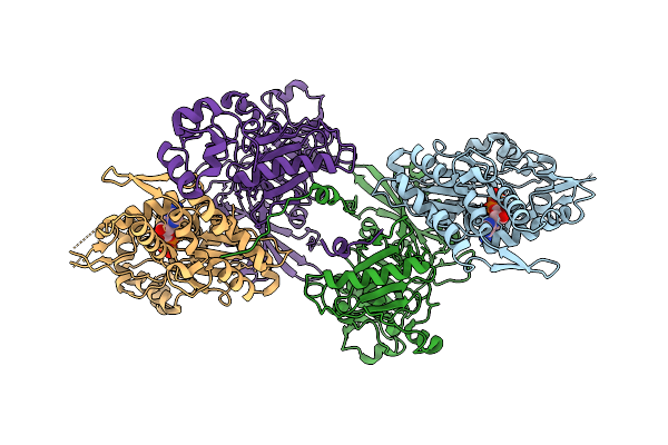
Organism: Rhodococcus sp. usk13, Synthetic construct
Method: X-RAY DIFFRACTION Resolution:2.45 Å Release Date: 2023-11-01 Classification: TRANSCRIPTION Ligands: SO4 |
 |
Organism: Rhodococcus sp. usk13, Rhodococcus
Method: X-RAY DIFFRACTION Resolution:2.96 Å Release Date: 2023-11-01 Classification: TRANSCRIPTION/DNA Ligands: PO4 |
 |
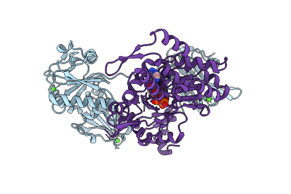
Organism: Rhodococcus sp. usk13
Method: X-RAY DIFFRACTION Resolution:1.44 Å Release Date: 2023-11-01 Classification: TRANSCRIPTION Ligands: CMP |
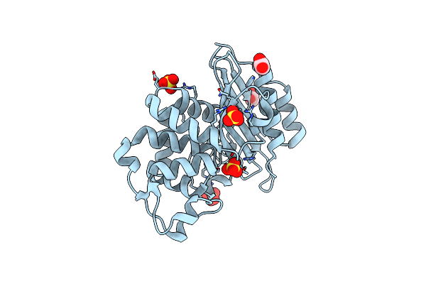 |
Organism: Micromonospora aurantiaca (strain atcc 27029 / dsm 43813 / bcrc 12538 / cbs 129.76 / jcm 10878 / nbrc 16125 / nrrl b-16091 / ina 9442)
Method: X-RAY DIFFRACTION Resolution:1.40 Å Release Date: 2020-04-22 Classification: HYDROLASE Ligands: ACT, SO4 |
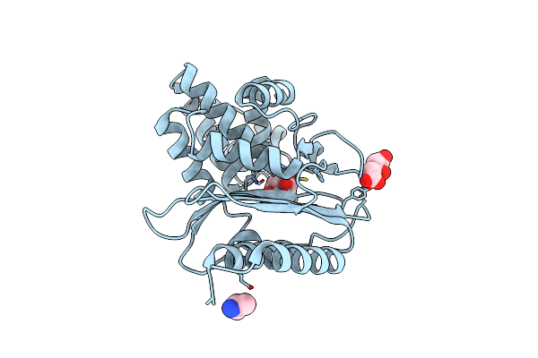 |
Organism: Desulfarculus baarsii (strain atcc 33931 / dsm 2075 / vkm b-1802 / 2st14)
Method: X-RAY DIFFRACTION Resolution:1.00 Å Release Date: 2020-04-22 Classification: HYDROLASE Ligands: ALA, CIT |
 |
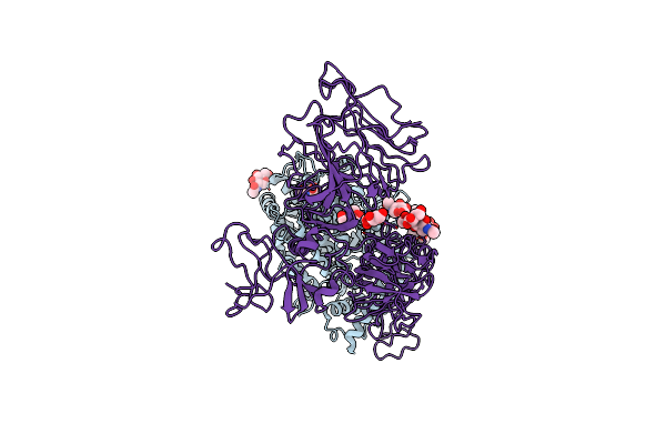
Organism: Komagataella phaffii (strain gs115 / atcc 20864), Homo sapiens, Komagataella phaffii gs115
Method: ELECTRON MICROSCOPY Release Date: 2020-02-12 Classification: TRANSCRIPTION |
 |
Organism: Komagataella phaffii (strain gs115 / atcc 20864), Komagataella phaffii gs115
Method: ELECTRON MICROSCOPY Release Date: 2020-01-29 Classification: TRANSCRIPTION |
 |
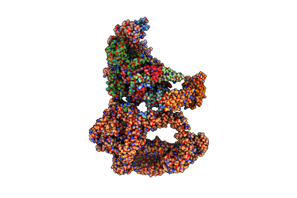
Rna Polymerase Ii Elongation Complex Bound With Elf1 And Spt4/5, Stalled At Shl(-1) Of The Nucleosome
Organism: Komagataella phaffii (strain gs115 / atcc 20864), Komagataella phaffii, Homo sapiens, Synthetic construct, Komagataella phaffii (strain atcc 76273 / cbs 7435 / cect 11047 / nrrl y-11430 / wegner 21-1)
Method: ELECTRON MICROSCOPY Release Date: 2019-02-20 Classification: TRANSCRIPTION/RNA/DNA Ligands: ZN, MG |
 |
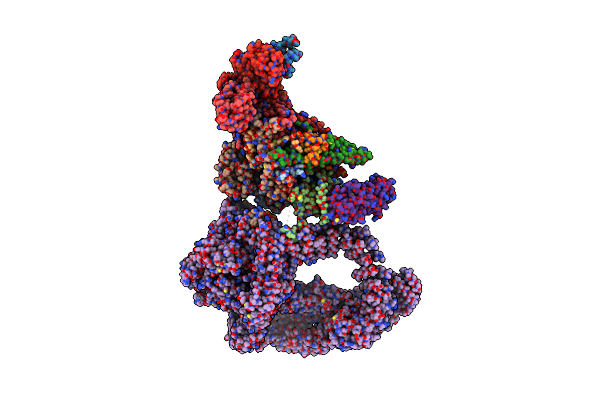
Rna Polymerase Ii Elongation Complex Bound With Elf1 And Spt4/5, Stalled At Shl(-5) Of The Nucleosome
Organism: Komagataella phaffii (strain gs115 / atcc 20864), Komagataella pastoris, Homo sapiens, Synthetic construct, Komagataella phaffii (strain atcc 76273 / cbs 7435 / cect 11047 / nrrl y-11430 / wegner 21-1)
Method: ELECTRON MICROSCOPY Release Date: 2019-02-20 Classification: TRANSCRIPTION/RNA/DNA Ligands: ZN, MG |