
Search Count: 643
 |
Organism: Stichodactyla helianthus
Method: ELECTRON MICROSCOPY Release Date: 2025-10-29 Classification: MEMBRANE PROTEIN Ligands: FO4, CLR |
 |
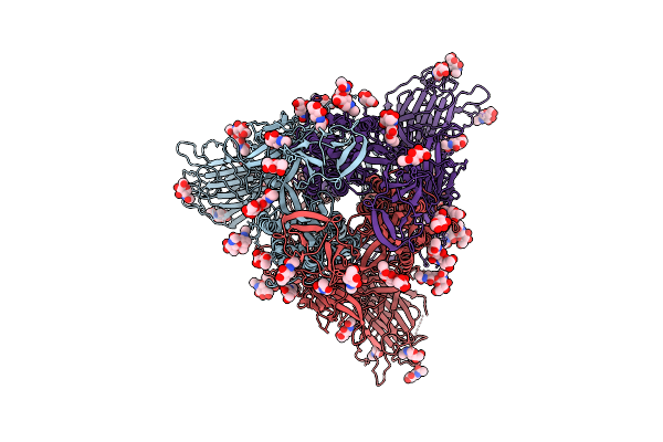
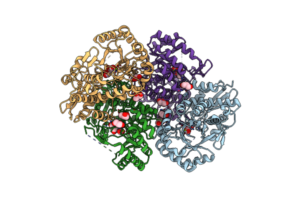
Structure Of 6Mer Pore Intermediate Of Sticholysin Ii (Stnii) Toxin In Lipid Nanodiscs
Organism: Stichodactyla helianthus
Method: ELECTRON MICROSCOPY Release Date: 2025-10-08 Classification: MEMBRANE PROTEIN Ligands: FO4 |
 |
Structure Of 5Mer Pore Intermediate Of Sticholysin Ii (Stnii) Toxin In Lipid Nanodiscs
Organism: Stichodactyla helianthus
Method: ELECTRON MICROSCOPY Release Date: 2025-09-17 Classification: MEMBRANE PROTEIN Ligands: FO4 |
 |
Organism: Bat sars-like coronavirus khosta-1
Method: ELECTRON MICROSCOPY Release Date: 2025-08-20 Classification: VIRAL PROTEIN Ligands: NAG |
 |
Crystal Structure Of Adenosylcobinamide Kinase / Adenosylcobinamide Phosphate Guanylyltransferase From Methylocapsa Palsarum
Organism: Methylocapsa palsarum
Method: X-RAY DIFFRACTION Resolution:1.80 Å Release Date: 2025-01-01 Classification: TRANSFERASE Ligands: PO4 |
 |
Crystal Structure Of Adenosylcobinamide Kinase / Adenosylcobinamide Phosphate Guanylyltransferase Complexed With Adenosylcobinamide-Phosphate
Organism: Methylocapsa palsarum
Method: X-RAY DIFFRACTION Resolution:2.60 Å Release Date: 2025-01-01 Classification: TRANSFERASE Ligands: B12, 5AD, PO4 |
 |
Adenosylcobinamide Kinase/Adenosylcobinamide Phosphate Guanylyltransferase -C91S
Organism: Methylocapsa palsarum
Method: X-RAY DIFFRACTION Resolution:1.50 Å Release Date: 2025-01-01 Classification: TRANSFERASE Ligands: PO4 |
 |
Crystal Structure Of Adenosylcobinamide Kinase / Adenosylcobinamide Phosphate Guanylyltransferase Complexed With Gdp
Organism: Methylocapsa palsarum
Method: X-RAY DIFFRACTION Resolution:1.92 Å Release Date: 2025-01-01 Classification: TRANSFERASE Ligands: GDP, DPO |
 |
Organism: Castilleja foliolosa
Method: X-RAY DIFFRACTION Resolution:1.90 Å Release Date: 2024-12-11 Classification: HYDROLASE Ligands: GOL |
 |
Ligand-Free Structure Of The Grpu Microcompartment Shell Protein From Pectobacterium Wasabiae
Organism: Pectobacterium wasabiae
Method: X-RAY DIFFRACTION Resolution:2.79 Å Release Date: 2014-07-30 Classification: ELECTRON TRANSPORT |
 |
Organism: Escherichia coli, Pectobacterium wasabiae wpp163, Thermus thermophilus, Streptomyces puniceus
Method: X-RAY DIFFRACTION Resolution:3.45 Å Release Date: 2014-07-09 Classification: RIBOSOME/ANTIBIOTIC Ligands: MG, ZN |
 |
Tracing The Evolution Of Angucyclinone Monooxygenases: Structural Determinants For C-12B Hydroxylation And Substrate Inhibition In Pgae
Organism: Streptomyces sp. pga64
Method: X-RAY DIFFRACTION Resolution:2.40 Å Release Date: 2013-06-12 Classification: OXIDOREDUCTASE Ligands: FAD, EDO, GOL, SO4, CL |
 |
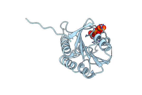
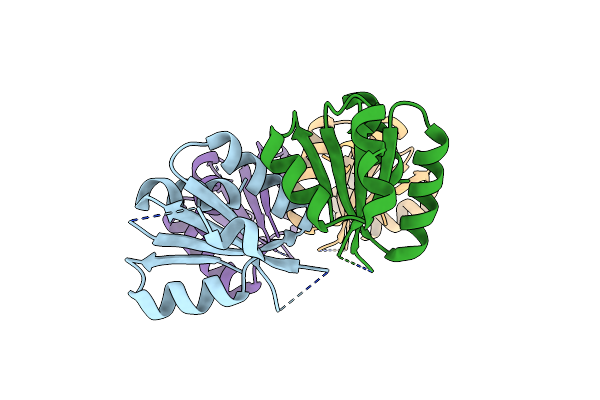
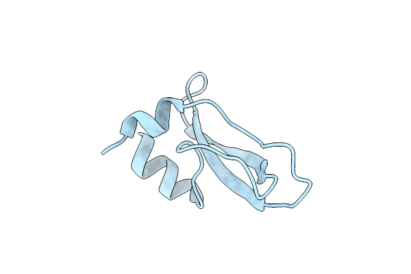
Crystal Structure Of The Kunitz-Type Protease Inhibitor Shpi-1 Lys13Leu Mutant In Complex With Pancreatic Elastase
Organism: Stichodactyla helianthus, Sus scrofa
Method: X-RAY DIFFRACTION Resolution:2.00 Å Release Date: 2012-11-21 Classification: HYDROLASE/Hydrolase Inhibitor Ligands: SO4, GOL |
 |
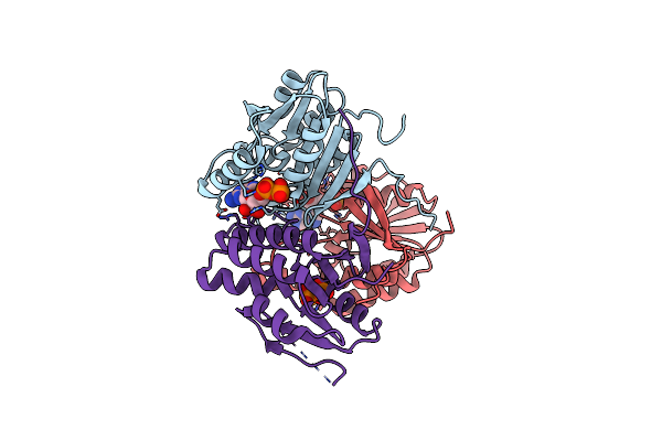
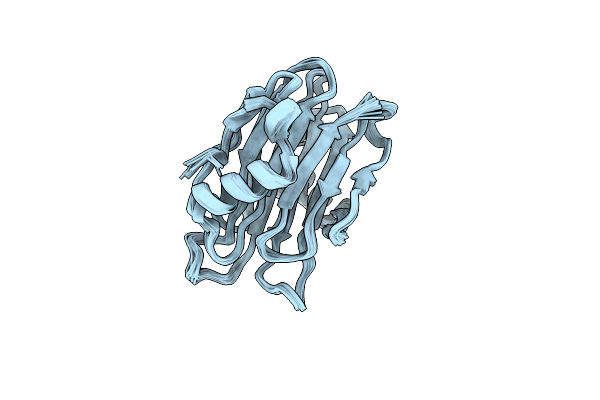
Crystal Structure Of Recombinant Kunitz Type Serine Protease Inhibitor-1 From The Caribbean Sea Anemone Stichodactyla Helianthus In Complex With Bovine Chymotrypsin
Organism: Stichodactyla helianthus, Bos taurus
Method: X-RAY DIFFRACTION Resolution:2.00 Å Release Date: 2012-08-01 Classification: HYDROLASE/HYDROLASE INHIBITOR Ligands: SO4 |
 |
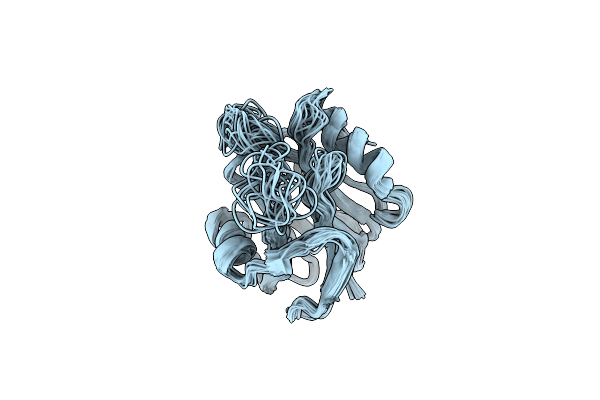
Crystal Structure Of Enolase Pc1_0802 (Target Efi-502240) From Pectobacterium Carotovorum Subsp. Carotovorum Pc1
Organism: Pectobacterium carotovorum subsp. carotovorum
Method: X-RAY DIFFRACTION Resolution:2.00 Å Release Date: 2012-03-28 Classification: ISOMERASE Ligands: GOL, CL, MG, FMT |
 |
Crystal Structure Of Recombinant Kunitz Type Serine Protease Inhibitor-1 From The Carribean Sea Anemone Stichodactyla Helianthus
Organism: Stichodactyla helianthus
Method: X-RAY DIFFRACTION Resolution:2.50 Å Release Date: 2011-08-17 Classification: HYDROLASE/HYDROLASE INHIBITOR Ligands: CL |
 |
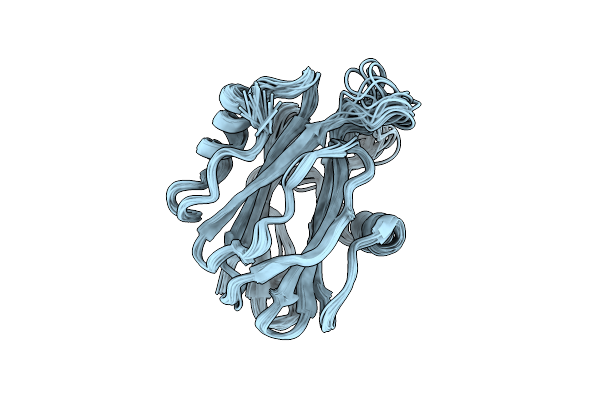
Structure Of Stnii-Y111N, A Mutant Of The Sea Anemone Actinoporin Sticholysin Ii
Organism: Stichodactyla helianthus
Method: SOLUTION NMR Release Date: 2011-06-29 Classification: TOXIN |
 |
Crystal Structure Of Recombinant Kunitz Type Serine Protease Inhibitor-1 From The Caribbean Sea Anemone Stichodactyla Helianthus In Complex With Bovine Pancreatic Trypsin
Organism: Stichodactyla helianthus, Bos taurus
Method: X-RAY DIFFRACTION Resolution:1.70 Å Release Date: 2011-03-16 Classification: Hydrolase/Hydrolase inhibitor Ligands: PO4 |
 |
Organism: Stichodactyla helianthus
Method: SOLUTION NMR Release Date: 2010-09-22 Classification: TOXIN |
 |
Organism: Stichodactyla helianthus
Method: SOLUTION NMR Release Date: 2010-09-01 Classification: TRANSPORT PROTEIN |