
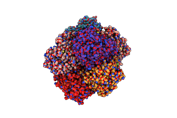
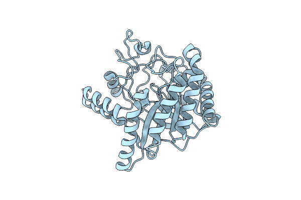
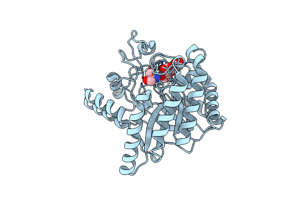
Search Count: 99
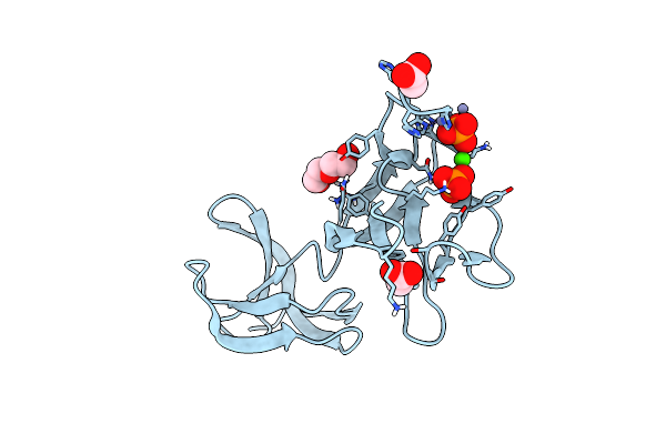 |
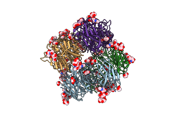
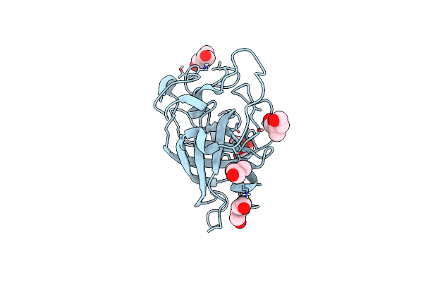
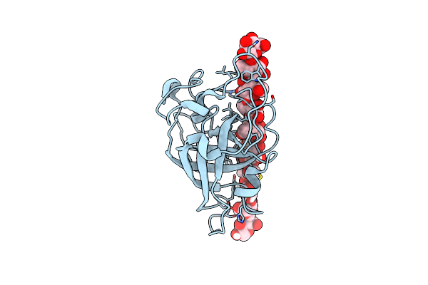
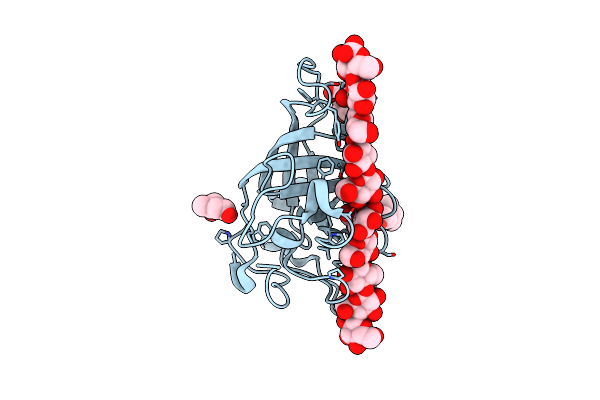
Crystal Structure Of S-Layer Protein Slpx From Lactobacillus Acidophilus, Domain Iii (Aa 363-499)
Organism: Lactobacillus acidophilus atcc 4796
Method: X-RAY DIFFRACTION Resolution:2.40 Å Release Date: 2023-08-23 Classification: CELL ADHESION Ligands: ZN, PO4, CA, ACT, PEG |
 |
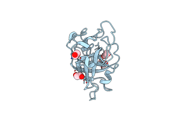
Organism: Phanerochaete chrysosporium
Method: X-RAY DIFFRACTION Resolution:2.54 Å Release Date: 2022-02-09 Classification: HYDROLASE Ligands: NAG, GOL |
 |
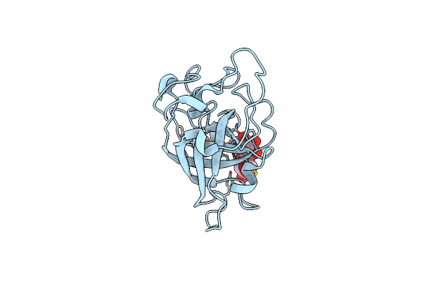
Organism: Phanerochaete chrysosporium
Method: X-RAY DIFFRACTION Resolution:3.08 Å Release Date: 2022-02-09 Classification: HYDROLASE Ligands: 1PE, PEG, EDO, NAG, XYP |
 |
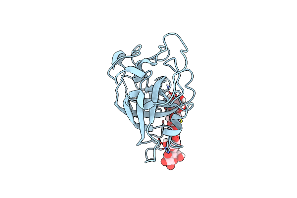
Crystal Structure Of Exo-Beta-1,3-Galactanase From Phanerochaete Chrysosporium Pc1,3Gal43A Apo Form
Organism: Phanerochaete chrysosporium
Method: X-RAY DIFFRACTION Resolution:1.40 Å Release Date: 2020-11-04 Classification: HYDROLASE Ligands: NAG, CA, CIT |
 |
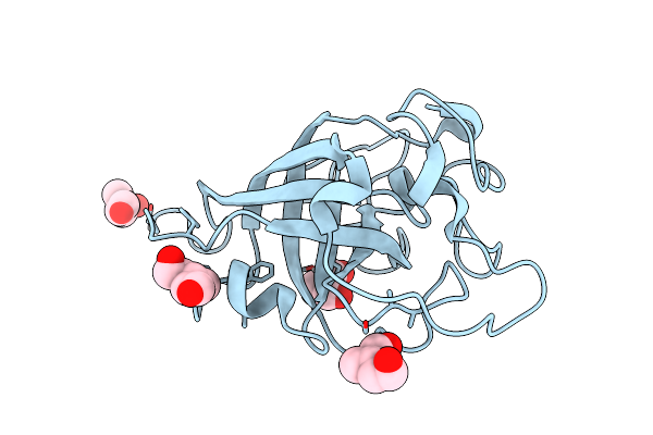
Crystal Structure Of Exo-Beta-1,3-Galactanase From Phanerochaete Chrysosporium Pc1,3Gal43A With Galactose
Organism: Phanerochaete chrysosporium
Method: X-RAY DIFFRACTION Resolution:1.50 Å Release Date: 2020-11-04 Classification: HYDROLASE Ligands: NAG, CA, GLA, GAL, ACY, ACT, GOL |
 |
Crystal Structure Of Exo-Beta-1,3-Galactanase From Phanerochaete Chrysosporium Pc1,3Gal43A E208Q With Beta-1,3-Galactotriose
Organism: Phanerochaete chrysosporium
Method: X-RAY DIFFRACTION Resolution:2.50 Å Release Date: 2020-11-04 Classification: HYDROLASE Ligands: NAG, CA, GAL |
 |
Crystal Structure Of Exo-Beta-1,3-Galactanase From Phanerochaete Chrysosporium Pc1,3Gal43A E208A With Beta-1,3-Galactotriose
Organism: Phanerochaete chrysosporium
Method: X-RAY DIFFRACTION Release Date: 2020-11-04 Classification: HYDROLASE Ligands: CA, ACT, GOL, NAG |
 |
Organism: Phanerochaete chrysosporium
Method: X-RAY DIFFRACTION Resolution:2.60 Å Release Date: 2018-10-10 Classification: OXIDOREDUCTASE Ligands: FAD, GOL |
 |
Organism: Phanerochaete chrysosporium
Method: X-RAY DIFFRACTION Resolution:2.50 Å Release Date: 2018-10-10 Classification: OXIDOREDUCTASE Ligands: FAD, GOL |
 |
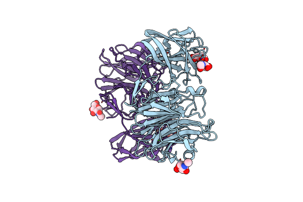
Structure Of The Cellobiohydrolase Cel6A From Phanerochaete Chrysosporium At 1.2 Angstrom
Organism: Phanerochaete chrysosporium
Method: X-RAY DIFFRACTION Resolution:1.20 Å Release Date: 2017-07-26 Classification: HYDROLASE |
 |
Structure Of The Cellobiohydrolase Cel6A From Phanerochaete Chrysosporium In Complex With Cellobiose At 2.1 Angstrom
Organism: Phanerochaete chrysosporium
Method: X-RAY DIFFRACTION Resolution:2.10 Å Release Date: 2017-07-26 Classification: HYDROLASE Ligands: TRS |
 |
Organism: Phanerochaete chrysosporium
Method: X-RAY DIFFRACTION Resolution:1.47 Å Release Date: 2017-06-21 Classification: HYDROLASE Ligands: EDO |
 |
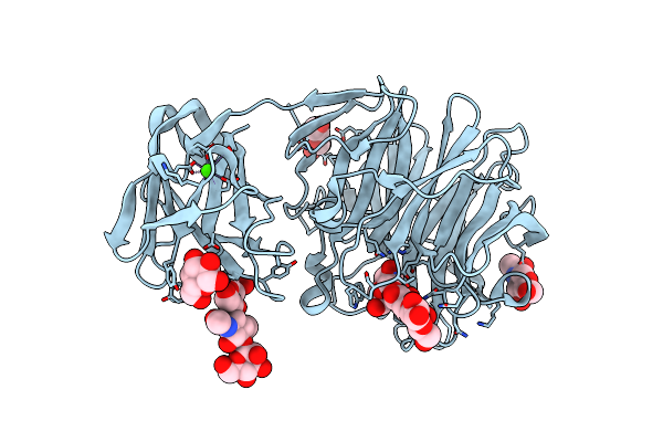
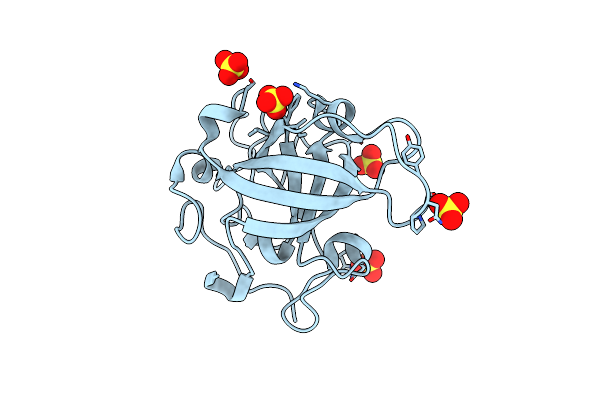
X-Ray Structure Of Pccel45A In Complex With Cellobiose Expressed In Aspergillus Nidullans
Organism: Phanerochaete chrysosporium
Method: X-RAY DIFFRACTION Resolution:1.70 Å Release Date: 2017-06-21 Classification: SUGAR BINDING PROTEIN Ligands: SO4 |
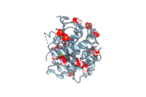 |
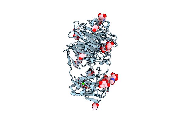
Ure2P5 From Phanerochaete Chrysosporium Cocrystallized With 1-(S-Glutathionyl)-2,4-Dinitrobenzene.
Organism: Phanerochaete chrysosporium
Method: X-RAY DIFFRACTION Resolution:2.03 Å Release Date: 2017-05-24 Classification: TRANSFERASE Ligands: GDS, NIN, SO4, GOL |
 |
Organism: Phanerochaete chrysosporium
Method: X-RAY DIFFRACTION Resolution:0.83 Å Release Date: 2015-10-14 Classification: HYDROLASE Ligands: 40S, TRS |
 |
X-Ray Structure Of Pccel45A With Cellopentaose At 0.64 Angstrom Resolution.
Organism: Phanerochaete chrysosporium
Method: X-RAY DIFFRACTION Resolution:0.64 Å Release Date: 2015-10-14 Classification: HYDROLASE |
 |
Organism: Phanerochaete chrysosporium
Method: X-RAY DIFFRACTION Resolution:1.20 Å Release Date: 2015-10-14 Classification: HYDROLASE Ligands: SO4 |
 |
Neutron And X-Ray Joint Refined Structure Of Pccel45A With Cellopentaose At 298K.
Organism: Phanerochaete chrysosporium
Method: NEUTRON DIFFRACTION, X-RAY DIFFRACTION Release Date: 2015-10-14 Classification: HYDROLASE Ligands: DOD |
 |
Organism: Phanerochaete chrysosporium
Method: X-RAY DIFFRACTION Resolution:1.00 Å Release Date: 2015-10-07 Classification: HYDROLASE Ligands: 40S, TRS |
 |
Organism: Phanerochaete chrysosporium
Method: X-RAY DIFFRACTION Resolution:0.99 Å Release Date: 2015-10-07 Classification: HYDROLASE Ligands: 40S |