
Search Count: 285
 |
Organism: Escherichia coli bl21
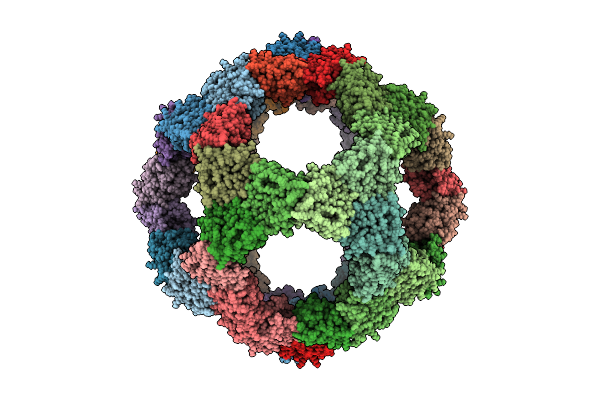
Method: ELECTRON MICROSCOPY Release Date: 2025-07-16 Classification: VIRUS LIKE PARTICLE |
 |
Organism: Escherichia coli bl21
Method: ELECTRON MICROSCOPY Release Date: 2025-03-12 Classification: LYASE |
 |
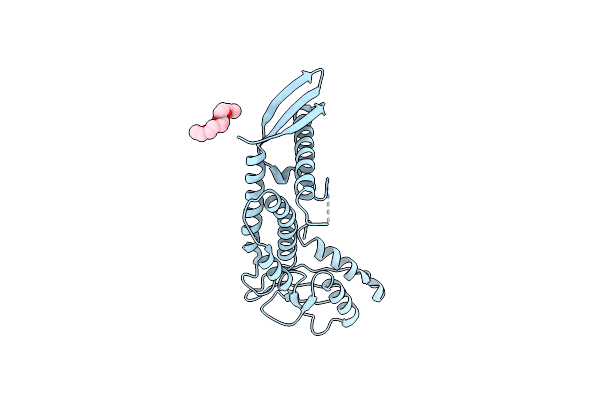
Cryo-Em Structure Of A Nautilus-Like Hflk/C Assembly In Complex With Ftsh Aaa Protease
Organism: Escherichia coli bl21
Method: ELECTRON MICROSCOPY Release Date: 2024-11-13 Classification: CHAPERONE |
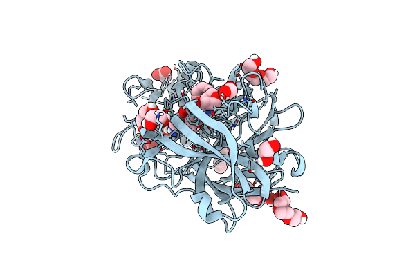 |
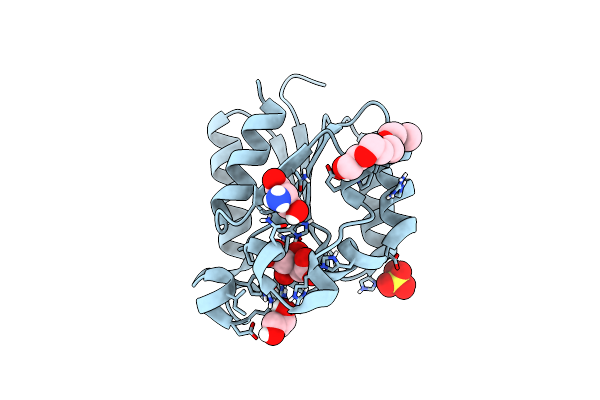
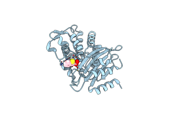
The Hiv Protease Inhibitor Amprenavir Binding To The Active Site Of Cryphonectria Parasitica Endothiapepsin
Organism: Cryphonectria parasitica
Method: X-RAY DIFFRACTION Resolution:1.80 Å Release Date: 2024-07-10 Classification: HYDROLASE Ligands: 478, PG4, PGE, EDO, ACY |
 |
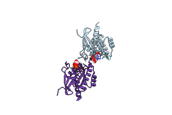
Rna Polymerase At U-Rich Pause Bound To Regulatory Rna Putl - Active, Closed Clamp State
Organism: Escherichia coli bl21, Escherichia coli k-12, Escherichia coli
Method: ELECTRON MICROSCOPY Release Date: 2022-10-19 Classification: TRANSCRIPTION Ligands: MG, ZN |
 |
Organism: Escherichia coli bl21, Synthetic construct
Method: X-RAY DIFFRACTION Resolution:2.10 Å Release Date: 2022-07-27 Classification: SIGNALING PROTEIN/TRANSCRIPTION Ligands: DHT, GOL |
 |
The Crystal Structure Of Type Ii Dehydroquinase From Butyrivibrio Crossotus Dsm 2876
Organism: Butyrivibrio crossotus dsm 2876
Method: X-RAY DIFFRACTION Resolution:1.70 Å Release Date: 2019-10-23 Classification: BIOSYNTHETIC PROTEIN Ligands: TLA, PO4, GOL |
 |
The Crystal Structure Of Type Ii Dehydroquinase From Butyrivibrio Crossotus Dsm 2876
Organism: Butyrivibrio crossotus dsm 2876
Method: X-RAY DIFFRACTION Resolution:1.05 Å Release Date: 2019-10-23 Classification: BIOSYNTHETIC PROTEIN Ligands: 3DS |
 |
The Crystal Structure Of Type Ii Dehydroquinase From Butyrivibrio Crossotus Dsm 2876
Organism: Butyrivibrio crossotus dsm 2876
Method: X-RAY DIFFRACTION Resolution:0.92 Å Release Date: 2019-10-23 Classification: BIOSYNTHETIC PROTEIN Ligands: ETE, SER, SO4, FLC, GOL |
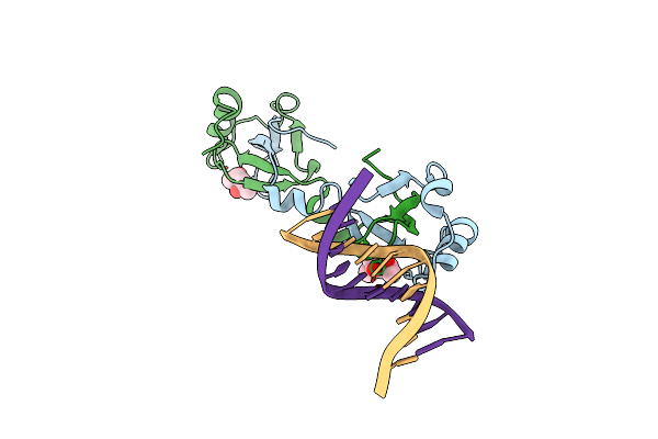 |
Crystal Structure Of Pwwp-Dna Complex For Human Hepatoma-Derived Growth Factor
Organism: Escherichia coli bl21, Rattus norvegicus
Method: X-RAY DIFFRACTION Resolution:2.84 Å Release Date: 2018-06-20 Classification: HORMONE Ligands: MPD |
 |
Organism: Cryphonectria parasitica, Synthetic construct
Method: X-RAY DIFFRACTION Resolution:2.89 Å Release Date: 2016-03-23 Classification: hydrolase/dna Ligands: CA, EDO |
 |
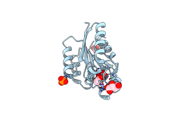
Neutron Structure Of A Perdeuterated Toho-1 R274N R276N Double Mutant Beta-Lactamase In Complex With A Fully Deuterated Boronic Acid (Bzb) At 100K
Organism: Escherichia coli
Method: NEUTRON DIFFRACTION Resolution:2.20 Å Release Date: 2014-07-09 Classification: HYDROLASE Ligands: BZB, DOD |
 |
Crystal Structure Of The Secreted Protein Hp1454 From The Human Pathogen Helicobacter Pylori
Organism: Helicobacter pylori r046wa
Method: X-RAY DIFFRACTION Resolution:2.70 Å Release Date: 2014-06-04 Classification: UNKNOWN FUNCTION Ligands: 1PE |
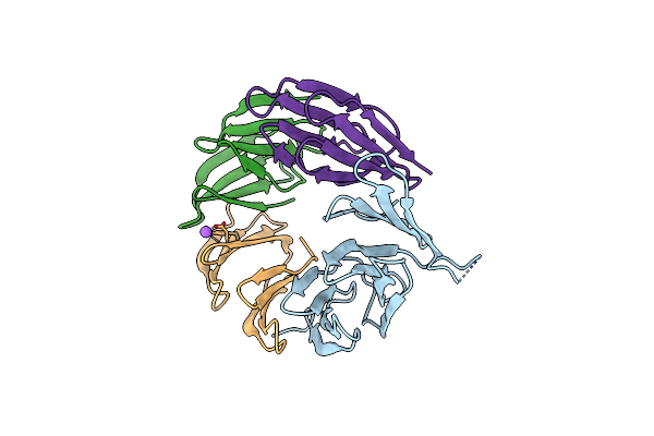 |
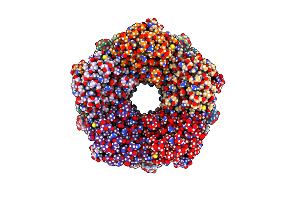
Structure And Assembly Of A B-Propeller With Nine Blades And A New Conserved Repetitive Sequence Motif
Organism: Escherichia coli
Method: X-RAY DIFFRACTION Resolution:2.10 Å Release Date: 2013-10-30 Classification: CHAPERONE |
 |
Visualizing Groel-Es In The Act Of Encapsulating A Non-Native Substrate Protein
Organism: Escherichia coli bl21, Escherichia coli k-12
Method: ELECTRON MICROSCOPY Resolution:8.90 Å Release Date: 2013-06-19 Classification: CHAPERONE Ligands: MG, ADP |
 |
Visualizing Groel-Es In The Act Of Encapsulating A Non-Native Substrate Protein
Organism: Escherichia coli bl21, Escherichia coli k-12
Method: ELECTRON MICROSCOPY Resolution:9.20 Å Release Date: 2013-06-19 Classification: PROTEIN FOLDING Ligands: ADP, MG |
 |
Visualizing Groel-Es In The Act Of Encapsulating A Non-Native Substrate Protein
Organism: Escherichia coli bl21, Escherichia coli k-12
Method: ELECTRON MICROSCOPY Resolution:15.90 Å Release Date: 2013-06-19 Classification: CHAPERONE Ligands: MG, ADP |
 |
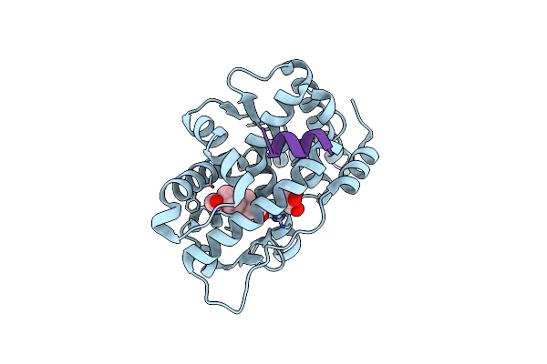
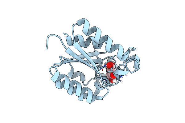
Crystal Structure Of A Ras-Like Protein From Cryphonectria Parasitica In Complex With Gdp
Organism: Cryphonectria parasitica
Method: X-RAY DIFFRACTION Resolution:1.60 Å Release Date: 2013-06-05 Classification: SIGNALING PROTEIN Ligands: GDP, MG |
 |
Fitting Result In The O-Layer Of The Subnanometer Structure Of The Bacterial Pkm101 Type Iv Secretion System Core Complex Digested With Elastase
Organism: Escherichia coli
Method: ELECTRON MICROSCOPY Resolution:8.50 Å Release Date: 2013-04-03 Classification: CELL ADHESION |
 |
Fitting Results In The I-Layer Of The Subnanometer Structure Of The Bacterial Pkm101 Type Iv Secretion System Core Complex Digested With Elastase
Organism: Escherichia coli
Method: ELECTRON MICROSCOPY Resolution:8.50 Å Release Date: 2013-04-03 Classification: CELL ADHESION |