
Search Count: 29
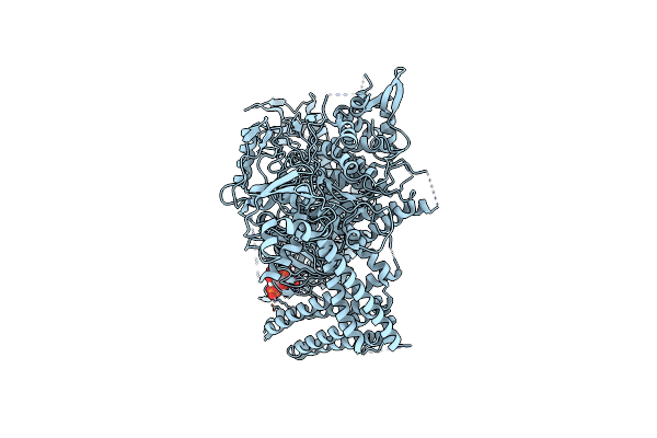 |
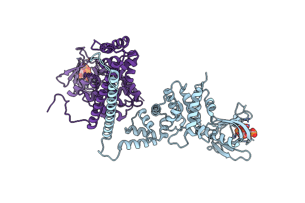
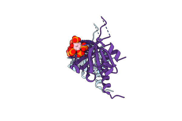
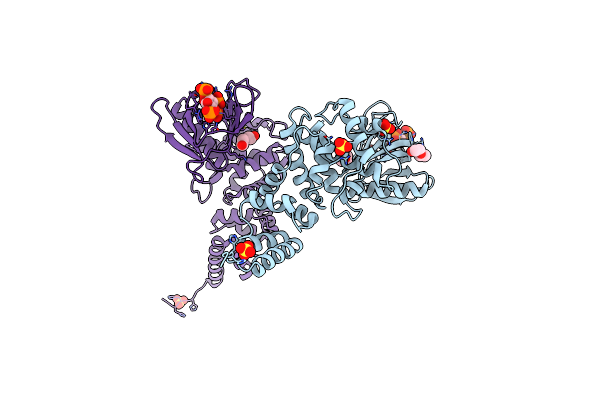
Full-Length P-Rex1 In Complex With Inositol 1,3,4,5-Tetrakisphosphate (Ip4)
Organism: Homo sapiens
Method: ELECTRON MICROSCOPY Release Date: 2024-04-10 Classification: SIGNALING PROTEIN Ligands: 4IP |
 |
Structure Of The Ptp-Like Myo-Inositol Phosphatase From Legionella Pneumophila Str. Paris In Complex With Myo-Inositol-(1,3,4,5)-Tetrakisphosphate
Organism: Legionella pneumophila str. paris
Method: X-RAY DIFFRACTION Resolution:1.85 Å Release Date: 2022-10-19 Classification: HYDROLASE Ligands: 4IP |
 |
Organism: Homo sapiens
Method: X-RAY DIFFRACTION Resolution:2.43 Å Release Date: 2021-04-07 Classification: CELL ADHESION Ligands: 4IP, EDO |
 |
Best Fitting Antiparallel Model For Volume 1 Of Truncated Dimeric Cytohesin-3 (Grp1; Amino Acids 14-399)
Organism: Homo sapiens
Method: ELECTRON MICROSCOPY Release Date: 2019-09-25 Classification: ENDOCYTOSIS Ligands: 4IP |
 |
Best Fitting Antiparallel Model For Volume 2 Of Truncated Dimeric Cytohesin-3 (Grp1; Amino Acids 14-399)
Organism: Homo sapiens
Method: ELECTRON MICROSCOPY Release Date: 2019-09-25 Classification: ENDOCYTOSIS Ligands: 4IP |
 |
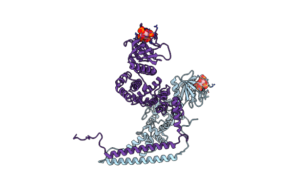
Model For Compact Volume Of Truncated Monomeric Cytohesin-3 (Grp1; Amino Acids 63-399) E161A 6Gs Arf6 Q67L Fusion Protein
Organism: Mus musculus, Homo sapiens
Method: ELECTRON MICROSCOPY Resolution:35.00 Å Release Date: 2018-01-10 Classification: LIPID BINDING PROTEIN Ligands: GTP, MG, 4IP |
 |
Model For Extended Volume Of Truncated Monomeric Cytohesin-3 (Grp1; Amino Acids 63-399) E161A Arf6 Q67L Fusion Protein
Organism: Mus musculus, Homo sapiens
Method: ELECTRON MICROSCOPY Release Date: 2018-01-10 Classification: LIPID BINDING PROTEIN Ligands: GTP, MG, 4IP |
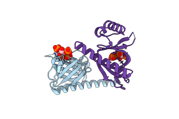 |
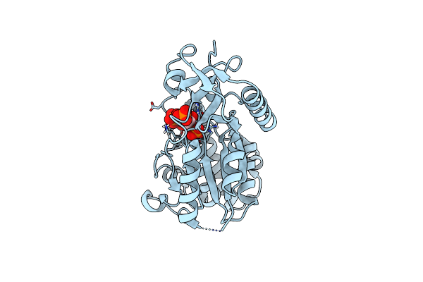
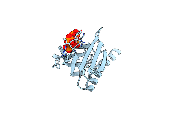
Crystal Structure Of The P-Rex1 Ph Domain With Inositol-(1,3,4,5)-Tetrakisphosphate Bound
Organism: Homo sapiens
Method: X-RAY DIFFRACTION Resolution:1.69 Å Release Date: 2016-04-20 Classification: LIPID BINDING PROTEIN Ligands: 4IP |
 |
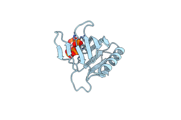
Crystal Structure Of The P-Rex1 Ph Domain With Inositol-(1,3,4,5)-Tetrakisphosphate Bound
Organism: Homo sapiens
Method: X-RAY DIFFRACTION Resolution:1.95 Å Release Date: 2016-04-20 Classification: LIPID BINDING PROTEIN Ligands: 4IP |
 |
Structure Of The Ptp-Like Myo-Inositol Phosphatase From Mitsuokella Multacida In Complex With Myo-Inositol-(1,3,4,5)-Tetrakisphosphate
Organism: Mitsuokella multacida
Method: X-RAY DIFFRACTION Resolution:2.20 Å Release Date: 2015-12-09 Classification: HYDROLASE Ligands: 4IP, PO4, GOL |
 |
Structure Of The Ptp-Like Myo-Inositol Phosphatase From Selenomonas Ruminantium In Complex With Myo-Inositol-(1,3,4,5)-Tetrakisphosphate
Organism: Selenomonas ruminantium
Method: X-RAY DIFFRACTION Resolution:2.10 Å Release Date: 2015-11-04 Classification: HYDROLASE Ligands: 4IP, GOL, CL |
 |
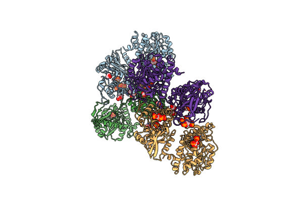
Organism: Homo sapiens
Method: X-RAY DIFFRACTION Resolution:1.85 Å Release Date: 2013-08-14 Classification: PROTEIN BINDING/SIGNALING PROTEIN Ligands: GTP, MG, CIT, GOL, K, 4IP |
 |
Structure Of The Ptp-Like Phytase From Selenomonas Ruminantium In Complex With Myo-Inositol (1,3,4,5)Tetrakisphosphate
Organism: Selenomonas ruminantium
Method: X-RAY DIFFRACTION Resolution:1.85 Å Release Date: 2011-12-07 Classification: HYDROLASE Ligands: ACT, GOL, 4IP, CL |
 |
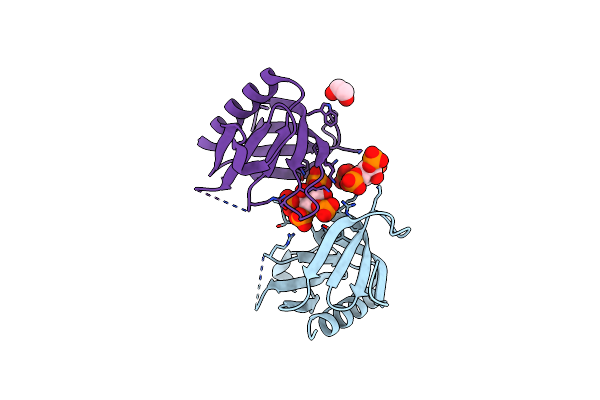
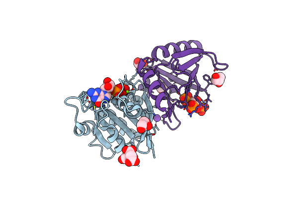
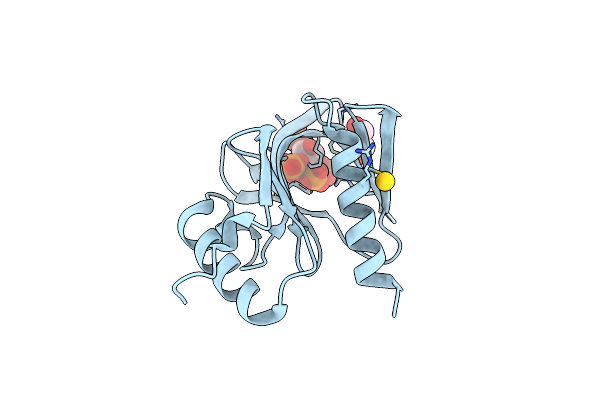
Structural Basis Of Phosphoinositide Binding To Kindlin-2 Pleckstrin Homology Domain In Regulating Integrin Activation
Organism: Homo sapiens
Method: SOLUTION NMR Release Date: 2011-10-26 Classification: CELL ADHESION Ligands: 4IP |
 |
Crystal Structure Of Programmed Cell Death 10 In Complex With Inositol 1,3,4,5-Tetrakisphosphate
Organism: Homo sapiens
Method: X-RAY DIFFRACTION Resolution:2.30 Å Release Date: 2010-06-30 Classification: APOPTOSIS Ligands: 4IP |
 |
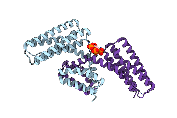
Organism: Mus musculus
Method: X-RAY DIFFRACTION Resolution:1.90 Å Release Date: 2007-12-04 Classification: SIGNALING PROTEIN Ligands: SO4, 4IP, PGE, PE5 |
 |
Organism: Mus musculus
Method: X-RAY DIFFRACTION Resolution:2.04 Å Release Date: 2007-12-04 Classification: SIGNALING PROTEIN Ligands: SO4, 4IP, PEG |
 |
A Transforming Mutation In The Pleckstrin Homology Domain Of Akt1 In Cancer (Akt1-Ph_E17K)
Organism: Homo sapiens
Method: X-RAY DIFFRACTION Resolution:2.46 Å Release Date: 2007-07-17 Classification: TRANSFERASE Ligands: 4IP |
 |
Triglycine Variant Of The Arno Pleckstrin Homology Domain In Complex With Ins(1,3,4,5)P4
Organism: Mus musculus
Method: X-RAY DIFFRACTION Resolution:2.30 Å Release Date: 2005-02-01 Classification: LIPID BINDING PROTEIN Ligands: 4IP |
 |
Crystal Structure Of The Pdk1 Pleckstrin Homology (Ph) Domain Bound To Inositol (1,3,4,5)-Tetrakisphosphate
Organism: Homo sapiens
Method: X-RAY DIFFRACTION Resolution:1.50 Å Release Date: 2004-11-19 Classification: TRANSFERASE Ligands: GOL, AU, 4IP |