Planned Maintenance: Some services may turn out to be unavailable from 15th January, 2026 to 16th January, 2026. We apologize for the inconvenience!

Planned Maintenance: Some services may turn out to be unavailable from 15th January, 2026 to 16th January, 2026. We apologize for the inconvenience!
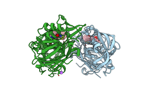 |
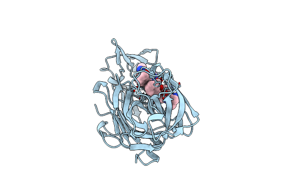
The Crystal Sturucture Of Rhizopuspepsin With A Bound Modified Peptide Inhibitor Generated By De Novo Drug Design.
Organism: Rhizopus microsporus var. chinensis, Synthetic construct
Method: X-RAY DIFFRACTION Resolution:1.21 Å Release Date: 2023-02-08 Classification: HYDROLASE |
 |
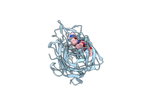
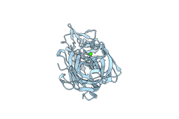
Organism: Rhizopus chinensis
Method: X-RAY DIFFRACTION Resolution:2.00 Å Release Date: 2019-10-09 Classification: HYDROLASE Ligands: SO4 |
 |
Organism: Plant transformation vector psiteii-4c1
Method: X-RAY DIFFRACTION Resolution:2.10 Å Release Date: 2011-08-03 Classification: FLUORESCENT PROTEIN Ligands: SO4, PE8 |
 |
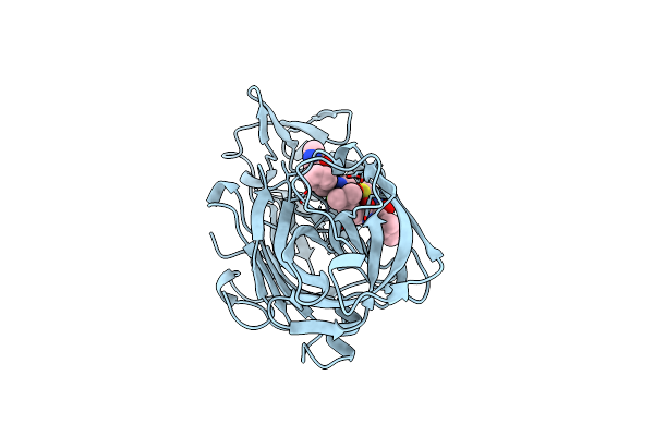
Structures Of Complexes Of Rhizopuspepsin With Pepstatin And Other Statine-Containing Inhibitors
Organism: Rhizopus chinensis
Method: X-RAY DIFFRACTION Resolution:2.50 Å Release Date: 1991-04-15 Classification: HYDROLASE/HYDROLASE INHIBITOR |
 |
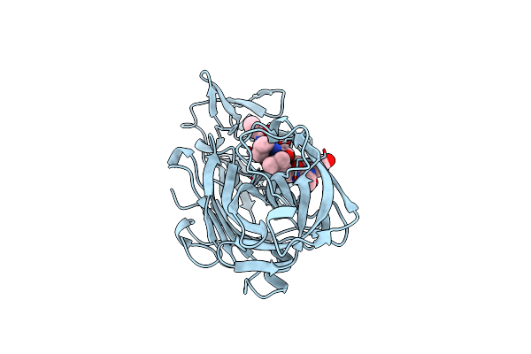
Structures Of Complexes Of Rhizopuspepsin With Pepstatin And Other Statine-Containing Inhibitors
Organism: Rhizopus chinensis
Method: X-RAY DIFFRACTION Resolution:2.10 Å Release Date: 1991-04-15 Classification: HYDROLASE/HYDROLASE INHIBITOR Ligands: CA |
 |
Structures Of Complexes Of Rhizopuspepsin With Pepstatin And Other Statine-Containing Inhibitors
Organism: Rhizopus chinensis, Streptomyces argenteolus subsp. toyonakensis
Method: X-RAY DIFFRACTION Resolution:2.50 Å Release Date: 1991-04-15 Classification: HYDROLASE/HYDROLASE INHIBITOR |
 |
Binding Of A Reduced Peptide Inhibitor To The Aspartic Proteinase From Rhizopus Chinensis. Implications For A Mechanism Of Action
Organism: Rhizopus microsporus var. chinensis
Method: X-RAY DIFFRACTION Resolution:1.80 Å Release Date: 1988-01-16 Classification: HYDROLASE/HYDROLASE INHIBITOR |
 |
Structure And Refinement At 1.8 Angstroms Resolution Of The Aspartic Proteinase From Rhizopus Chinensis
Organism: Rhizopus microsporus var. chinensis
Method: X-RAY DIFFRACTION Resolution:1.80 Å Release Date: 1987-07-16 Classification: HYDROLASE (ASPARTIC PROTEINASE) Ligands: CA |
 |
 |
 |
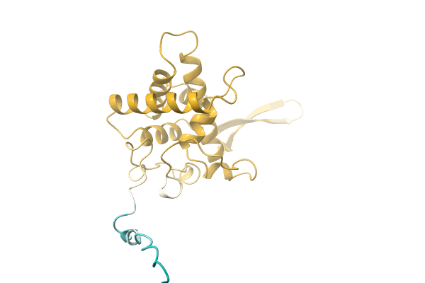
Organism: Prasinocyma nr. baumgaertneri EO-2018
Method: Alphafold Release Date: Classification: NA Ligands: NA |
 |
 |
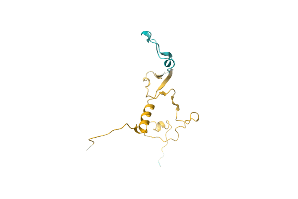
Organism: Oedicentra nr. albipennis EO-2018
Method: Alphafold Release Date: Classification: NA Ligands: NA |
 |
 |
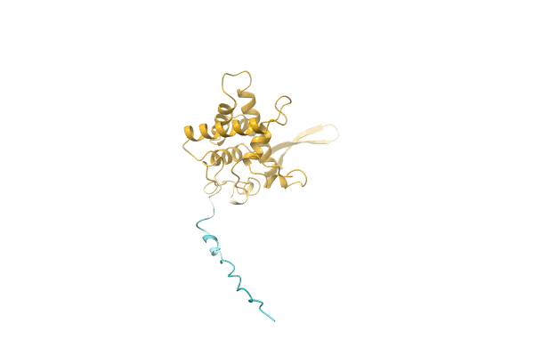
Organism: Asthenotricha nr. inutilis EO-2018
Method: Alphafold Release Date: Classification: NA Ligands: NA |
 |
 |
 |
 |
Organism: Comibaena nr. flavitaenia EO-2018
Method: Alphafold Release Date: Classification: NA Ligands: NA |
 |