
Search Count: 666
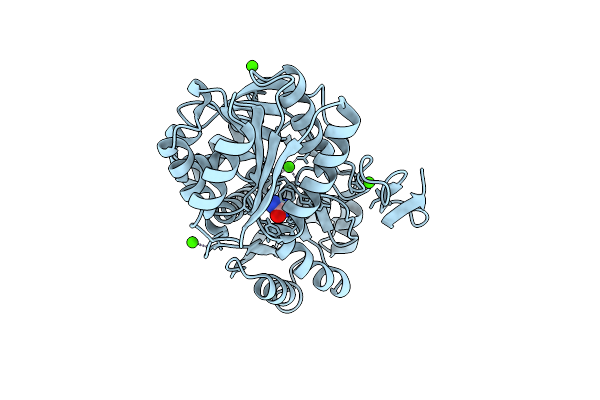 |
Crystal Structure Of Urta From Prochlorococcus Marinus Str. Mit 9313 In Complex With Urea And Calcium
Organism: Prochlorococcus marinus str. mit 9313
Method: X-RAY DIFFRACTION Resolution:1.60 Å Release Date: 2023-11-22 Classification: TRANSPORT PROTEIN Ligands: URE, CA |
 |
Solution Nmr Structure Of Prochlorosin 2.11 (Pcn2.11) Produced By Prochlorococcus Mit 9313
Organism: Prochlorococcus marinus str. mit 9313
Method: SOLUTION NMR Release Date: 2020-09-09 Classification: UNKNOWN FUNCTION |
 |
Solution Nmr Structure Of Prochlorosin 2.10 Produced By Prochlorococcus Mit 9313
Organism: Prochlorococcus marinus str. mit 9313
Method: SOLUTION NMR Release Date: 2020-09-09 Classification: UNKNOWN FUNCTION |
 |
Organism: Prochlorococcus marinus str. mit 9313
Method: SOLUTION NMR Release Date: 2020-07-08 Classification: UNKNOWN FUNCTION |
 |
Solution Nmr Structure Of Prochlorosin 2.1 Produced By Prochlorococcus Mit 9313
Organism: Prochlorococcus marinus str. mit 9313
Method: SOLUTION NMR Release Date: 2020-07-08 Classification: UNKNOWN FUNCTION |
 |
Organism: Prochlorococcus marinus str. mit 9313
Method: SOLUTION NMR Release Date: 2020-07-08 Classification: UNKNOWN FUNCTION |
 |
Crystal Structure Of A Double Glycine Motif Protease From Ams/Pcat Transporter In Complex With The Leader Peptide
Organism: Lachnospiraceae bacterium c6a11, Prochlorococcus marinus str. mit 9313
Method: X-RAY DIFFRACTION Resolution:2.00 Å Release Date: 2019-02-06 Classification: TRANSPORT PROTEIN Ligands: 16P |
 |
Organism: Salmonella phage det7
Method: X-RAY DIFFRACTION Resolution:1.63 Å Release Date: 2018-12-19 Classification: SUGAR BINDING PROTEIN Ligands: EDO |
 |
Organism: Salmonella phage det7
Method: X-RAY DIFFRACTION Resolution:2.10 Å Release Date: 2018-12-19 Classification: SUGAR BINDING PROTEIN Ligands: EDO |
 |
Insights Into Substrate And Metal Binding From The Crystal Structure Of Cyanobacterial Aldehyde Deformylating Oxygenase With Substrate Analogs Bound
Organism: Prochlorococcus marinus
Method: X-RAY DIFFRACTION Resolution:2.08 Å Release Date: 2015-02-11 Classification: OXIDOREDUCTASE Ligands: FE, Y69, EDO |
 |
Insights Into Substrate And Metal Binding From The Crystal Structure Of Cyanobacterial Aldehyde Deformylating Oxygenase With Substrate Bound
Organism: Prochlorococcus marinus
Method: X-RAY DIFFRACTION Resolution:2.17 Å Release Date: 2014-12-24 Classification: OXIDOREDUCTASE Ligands: FE, Y69 |
 |
Insights Into Substrate And Metal Binding From The Crystal Structure Of Cyanobacterial Aldehyde Deformylating Oxygenase With Substrate Bound
Organism: Prochlorococcus marinus
Method: X-RAY DIFFRACTION Resolution:1.90 Å Release Date: 2014-11-26 Classification: OXIDOREDUCTASE Ligands: FE, Y39, DMS |
 |
Insights Into Substrate And Metal Binding From The Crystal Structure Of Cyanobacterial Aldehyde Deformylating Oxygenase With Substrate Bound
Organism: Prochlorococcus marinus
Method: X-RAY DIFFRACTION Resolution:1.70 Å Release Date: 2014-11-19 Classification: OXIDOREDUCTASE Ligands: FE, Y39, DMS |
 |
Insights Into Substrate And Metal Binding From The Crystal Structure Of Cyanobacterial Aldehyde Deformylating Oxygenase With Substrate Bound
Organism: Prochlorococcus marinus
Method: X-RAY DIFFRACTION Resolution:1.60 Å Release Date: 2014-10-15 Classification: OXIDOREDUCTASE Ligands: FE, STE, EDO |
 |
Organism: Prochlorococcus marinus
Method: X-RAY DIFFRACTION Resolution:1.90 Å Release Date: 2014-08-27 Classification: STRUCTURAL PROTEIN |
 |
Crystal Structure Of A Niemann-Pick Type C2 Protein From Japanese Carpenter Ant
Organism: Camponotus japonicus
Method: X-RAY DIFFRACTION Resolution:1.80 Å Release Date: 2014-02-05 Classification: LIPID BINDING PROTEIN |
 |
Crystal Structure Of A Niemann-Pick Type C2 Protein From Japanese Carpenter Ant In Complex With Oleic Acid
Organism: Camponotus japonicus
Method: X-RAY DIFFRACTION Resolution:1.70 Å Release Date: 2014-02-05 Classification: LIPID BINDING PROTEIN Ligands: OLA |
 |
Crystal Structure Of Prochlorococcus Marinus Aldehyde-Deformylating Oxygenase Wild Type With Palmitic Acid Bound
Organism: Prochlorococcus marinus
Method: X-RAY DIFFRACTION Resolution:1.84 Å Release Date: 2013-06-26 Classification: OXIDOREDUCTASE Ligands: FE, PLM |
 |
Crystal Structure Of Prochlorococcus Marinus Aldehyde-Deformylating Oxygenase (Mutant V41Y)
Organism: Prochlorococcus marinus
Method: X-RAY DIFFRACTION Resolution:1.88 Å Release Date: 2013-06-26 Classification: OXIDOREDUCTASE Ligands: 6NA, FE |
 |
Crystal Structure Of Prochlorococcus Marinus Aldehyde-Deformylating Oxygenase (Mutant A134F)
Organism: Prochlorococcus marinus
Method: X-RAY DIFFRACTION Resolution:1.67 Å Release Date: 2013-06-26 Classification: OXIDOREDUCTASE Ligands: 6NA, FE |