
Search Count: 81
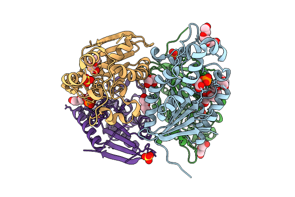 |
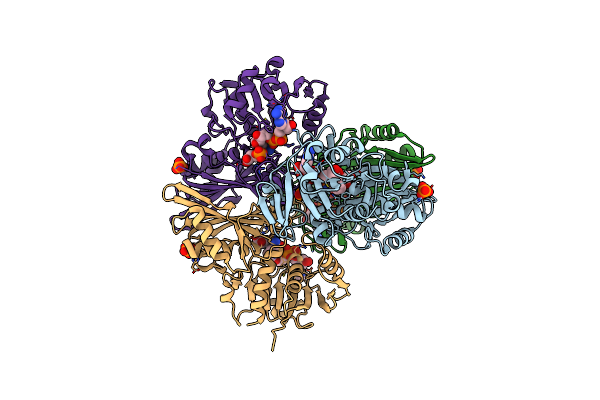
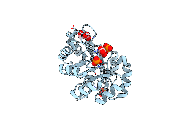
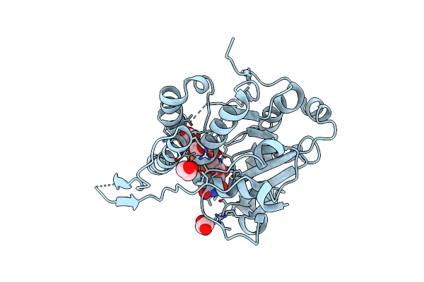
Crystal Structure Of Phosphoglycerate Mutase From Trichomonas Vaginalis In Complex With 3-Phosphoglyceric Acid
Organism: Trichomonas vaginalis g3
Method: X-RAY DIFFRACTION Resolution:2.10 Å Release Date: 2025-12-03 Classification: ISOMERASE Ligands: CL, 3PG, PG4, SO4 |
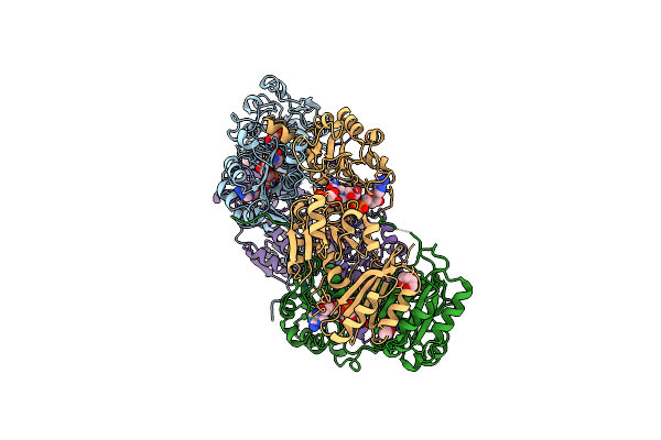 |
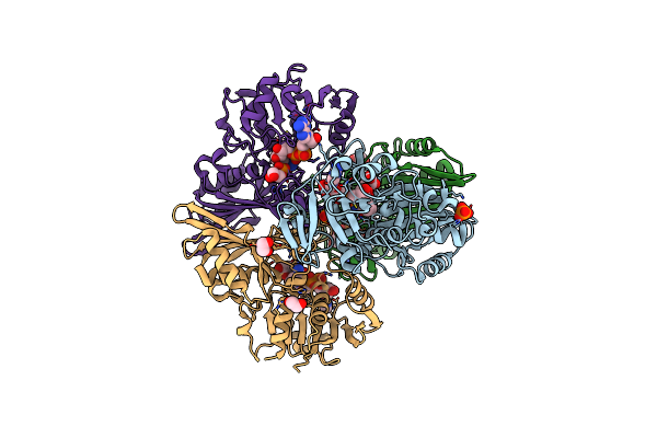
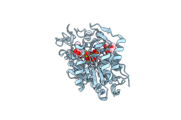
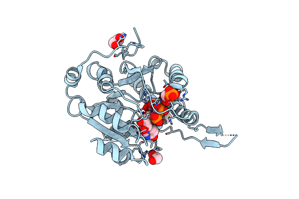
Organism: Saccharomyces cerevisiae
Method: X-RAY DIFFRACTION Resolution:2.70 Å Release Date: 2025-01-15 Classification: CYTOSOLIC PROTEIN Ligands: NAD, 3PG, BU1 |
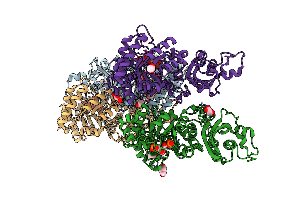 |
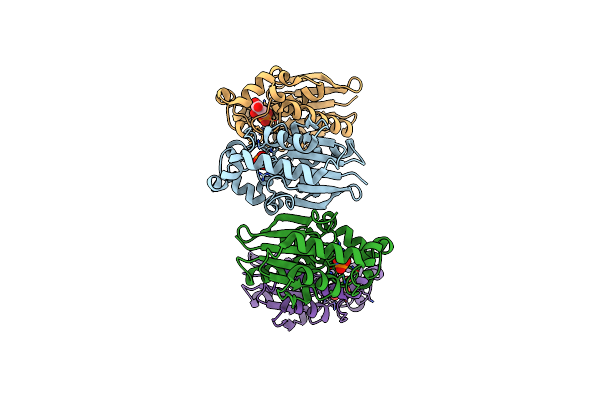
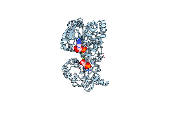
Organism: Archaeoglobus fulgidus
Method: X-RAY DIFFRACTION Resolution:1.70 Å Release Date: 2022-11-30 Classification: LYASE Ligands: MG, 3PG, GOL, ACT |
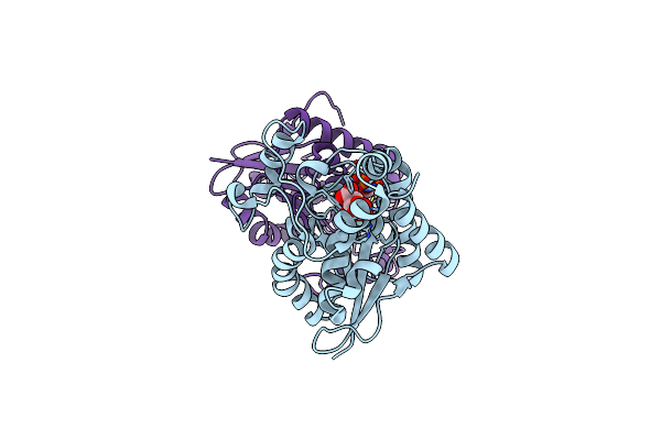 |
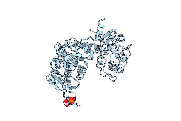
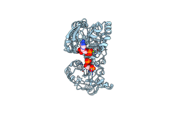
The Ternary Complex Of Human Bisphosphoglycerate Mutase With 3-Phosphoglycerate And 2-Phosphoglycolate
Organism: Homo sapiens
Method: X-RAY DIFFRACTION Resolution:2.25 Å Release Date: 2022-04-13 Classification: ISOMERASE Ligands: 3PG, PGA |
 |
Crystal Structure Of C150S Mutant Of Glyceraldehyde-3-Phosphate-Dehydrogenase1 From Escherichia Coli Complexed With G3P At 2.69 Angstrom Resolution
Organism: Escherichia coli bl21(de3)
Method: X-RAY DIFFRACTION Resolution:2.68 Å Release Date: 2021-05-26 Classification: OXIDOREDUCTASE Ligands: NAD, G3H, PO4, 3PG |
 |
Crystal Structure Of C150A+H177A Mutant Of Glyceraldehyde-3-Phosphate-Dehydrogenase1 From Escherichia Coli Complexed With G3P At 1.8 Angstrom Resolution
Organism: Escherichia coli bl21(de3)
Method: X-RAY DIFFRACTION Resolution:1.80 Å Release Date: 2021-05-26 Classification: OXIDOREDUCTASE Ligands: NAD, 3PG, PO4, G3H, EDO |
 |
Crystal Structure Of Phosphoserine Phosphatase In Complex With 3-Phosphoglyceric Acid From Entamoeba Histolytica
Organism: Entamoeba histolytica
Method: X-RAY DIFFRACTION Resolution:2.48 Å Release Date: 2021-03-03 Classification: CYTOSOLIC PROTEIN Ligands: PO4, 3PG |
 |
Crystal Structure Phgdh In Complex With Nadh And 3-Phosphoglycerate At 1.77 A Resolution
Organism: Homo sapiens
Method: X-RAY DIFFRACTION Resolution:1.77 Å Release Date: 2019-08-07 Classification: OXIDOREDUCTASE Ligands: 3PG, NAI |
 |
Crystal Structure Of Psychrophilic Phosphoglycerate Kinase From Pseudomonas Tacii18 In Complex With 3-Phosphoglycerate
Organism: Pseudomonas sp. 'tac ii 18'
Method: X-RAY DIFFRACTION Resolution:2.10 Å Release Date: 2019-06-12 Classification: TRANSFERASE Ligands: 3PG |
 |
Crystal Structure Of The 2-Keto-3-Deoxy-6-Phosphogluconate Aldolase Of Zymomonas Mobilis Zm4 With 3-Phosphoglycerate
Organism: Zymomonas mobilis subsp. mobilis (strain atcc 31821 / zm4 / cp4)
Method: X-RAY DIFFRACTION Resolution:1.96 Å Release Date: 2018-06-20 Classification: LYASE Ligands: PO4, 3PG, 3PY |
 |
Pgk1 In Complex With Crt0063465 (3-[2-(4-Bromophenyl)-5,7-Dimethyl-Pyrazolo[1,5-A]Pyrimidin-6-Yl]Propanoic Acid)
Organism: Homo sapiens
Method: X-RAY DIFFRACTION Resolution:1.90 Å Release Date: 2018-05-16 Classification: TRANSFERASE Ligands: 93T, EDO, 3PG |
 |
Organism: Homo sapiens
Method: X-RAY DIFFRACTION Resolution:2.05 Å Release Date: 2018-02-14 Classification: TRANSFERASE Ligands: MG, 3PG, ADP, BTB |
 |
Organism: Homo sapiens
Method: X-RAY DIFFRACTION Resolution:1.81 Å Release Date: 2017-12-20 Classification: TRANSFERASE Ligands: ADP, 3PG, MG |
 |
The X-Ray Structure Of Human M189I Pgk-1 Mutant In Partially Closed Conformation
Organism: Homo sapiens
Method: X-RAY DIFFRACTION Resolution:1.67 Å Release Date: 2017-11-29 Classification: TRANSFERASE Ligands: 3PG, PO4, ADP, MG |
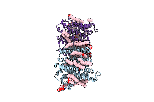 |
Crystal Structure Of The Triose-Phosphate/Phosphate Translocator In Complex With 3-Phosphoglycerate
Organism: Galdieria sulphuraria
Method: X-RAY DIFFRACTION Resolution:2.20 Å Release Date: 2017-10-04 Classification: TRANSPORT PROTEIN Ligands: OLC, 3PG, FLC |
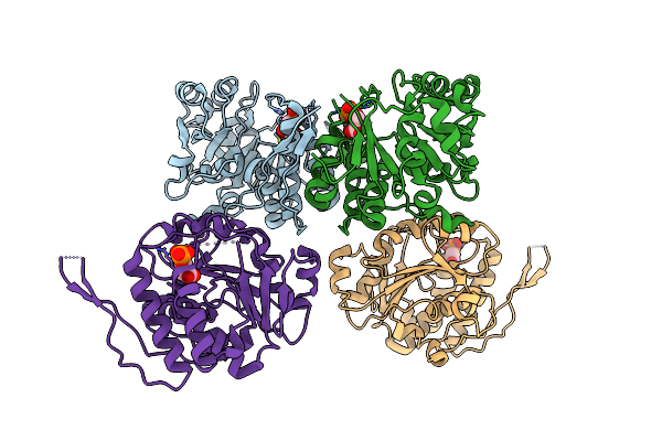 |
Crystal Structure Of Glucosyl-3-Phosphoglycerate Synthase From Mycobacterium Tuberculosis In Complex With Phosphoglyceric Acid (Pga) - Gpgs*Pga
Organism: Mycobacterium tuberculosis h37rv
Method: X-RAY DIFFRACTION Resolution:2.82 Å Release Date: 2017-05-24 Classification: TRANSFERASE Ligands: 3PG |
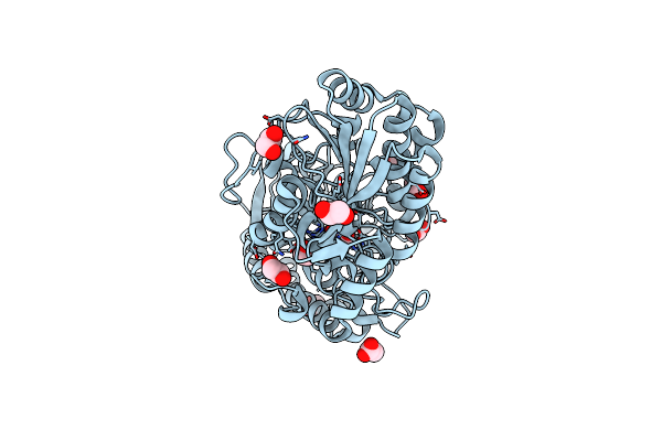 |
Crystal Structure Of 2,3-Bisphosphoglycerate-Independent Phosphoglycerate Mutase Bound To 3-Phosphoglycerate, From Acinetobacter Baumannii
Organism: Acinetobacter baumannii
Method: X-RAY DIFFRACTION Resolution:1.50 Å Release Date: 2017-05-24 Classification: ISOMERASE Ligands: MN, 3PG, EDO, ACT |
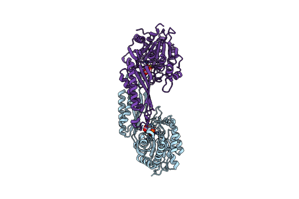 |
Crystal Structure Of Amidase, Hydantoinase/Carbamoylase Family From Burkholderia Vietnamiensis
Organism: Burkholderia vietnamiensis (strain g4 / lmg 22486)
Method: X-RAY DIFFRACTION Resolution:1.80 Å Release Date: 2016-03-02 Classification: HYDROLASE Ligands: ZN, 3PG |
 |
Crystal Structure Of Glucosyl-3-Phosphoglycerate Synthase From Mycobacterium Tuberculosis In Complex With Mn2+, Uridine-Diphosphate-Glucose (Udp-Glc) And Phosphoglyceric Acid (Pga) - Gpgs Mn2+ Udp-Glc Pga-1
Organism: Mycobacterium tuberculosis
Method: X-RAY DIFFRACTION Resolution:2.35 Å Release Date: 2015-07-15 Classification: TRANSFERASE Ligands: MN, 3PG, UPG, EDO |
 |
Organism: Mycobacterium tuberculosis (strain atcc 25618 / h37rv)
Method: X-RAY DIFFRACTION Resolution:2.27 Å Release Date: 2015-07-15 Classification: TRANSFERASE Ligands: MN, CL, 3PG, UPG, EDO |