
Search Count: 319
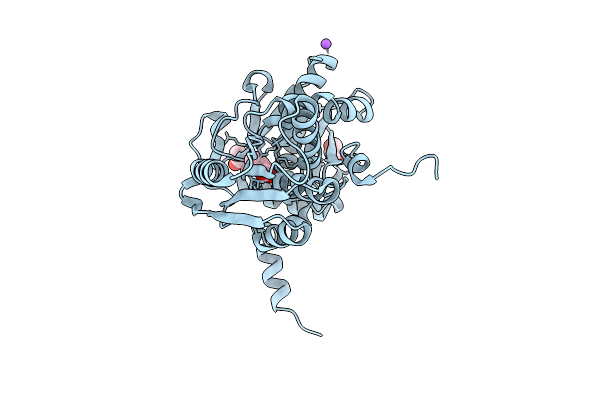 |
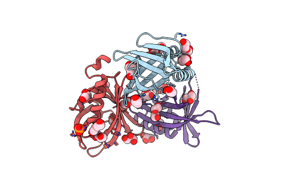
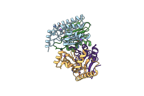
S-Methylcysteine Synthase (Bsas4) From Common Bean In Complex With Plp, Bez, And Gol
Organism: Phaseolus vulgaris
Method: X-RAY DIFFRACTION Resolution:1.60 Å Release Date: 2025-12-03 Classification: TRANSFERASE Ligands: BEZ, CL, GOL, NA |
 |
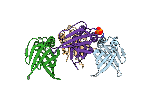
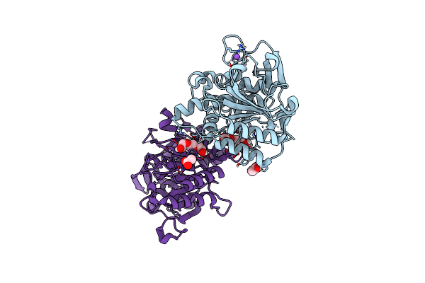
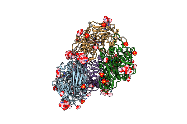
Crystal Structure Of Potassium-Independent L-Asparaginase From Phaseolus Vulgaris (Pvaiii, Pvaspg2)
Organism: Phaseolus vulgaris
Method: X-RAY DIFFRACTION Resolution:1.88 Å Release Date: 2025-05-14 Classification: HYDROLASE |
 |
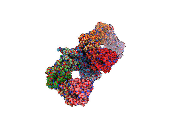
Organism: Phaseolus vulgaris, Brome mosaic virus
Method: ELECTRON MICROSCOPY Release Date: 2025-03-12 Classification: LIGASE/RNA Ligands: ATP |
 |
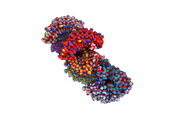
Organism: Phaseolus vulgaris, Brome mosaic virus
Method: ELECTRON MICROSCOPY Release Date: 2025-03-12 Classification: LIGASE/RNA |
 |
Organism: Phaseolus vulgaris, Brome mosaic virus
Method: ELECTRON MICROSCOPY Release Date: 2025-03-12 Classification: LIGASE/RNA |
 |
Organism: Phaseolus vulgaris, Fusarium phyllophilum
Method: X-RAY DIFFRACTION Resolution:1.94 Å Release Date: 2024-02-07 Classification: HYDROLASE |
 |
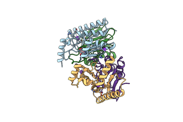
Trna-Like Structure From Brome Mosaic Virus Bound To Tyrosyl-Trna Synthetase From Phaseolus Vulgaris. Conformation: Bound State 1.
Organism: Phaseolus vulgaris, Brome mosaic virus
Method: ELECTRON MICROSCOPY Release Date: 2021-12-01 Classification: RNA BINDING PROTEIN/RNA |
 |
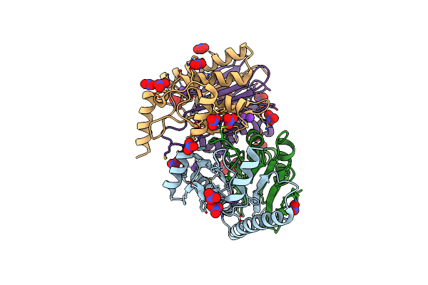
Trna-Like Structure From Brome Mosaic Virus Bound To Tyrosyl-Trna Synthetase From Phaseolus Vulgaris. Conformation: Bound State 2.
Organism: Phaseolus vulgaris, Brome mosaic virus
Method: ELECTRON MICROSCOPY Release Date: 2021-12-01 Classification: RNA BINDING PROTEIN/RNA |
 |
Mutations In Ssgb Correlate To Longitudinal Cell Division During Sporulation Of Streptomyces Coelicolor
Organism: Streptomyces sp. ag82_o1-9
Method: X-RAY DIFFRACTION Resolution:3.20 Å Release Date: 2020-09-30 Classification: CELL CYCLE Ligands: PO4 |
 |
Mutations In Ssgb Correlate To Longitudinal Cell Division During Sporulation Of Streptomyces Coelicolor
Organism: Streptomyces sp. ag82_o1-9
Method: X-RAY DIFFRACTION Resolution:2.30 Å Release Date: 2020-08-26 Classification: CELL CYCLE Ligands: PEG, GOL, PO4 |
 |
The Lipy/F-Motif In An Intracellular Subtilisin Protease Is Involved In Inhibition
Organism: Planococcus plakortidis
Method: X-RAY DIFFRACTION Resolution:1.30 Å Release Date: 2018-06-27 Classification: HYDROLASE Ligands: PGE, NA, ACT |
 |
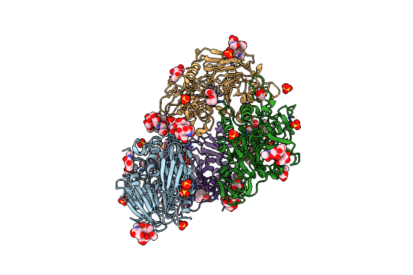
Crystal Structure Of Potassium-Dependent Plant-Type L-Asparaginase From Phaseolus Vulgaris In Complex With K+ Cations
Organism: Phaseolus vulgaris
Method: X-RAY DIFFRACTION Resolution:2.30 Å Release Date: 2014-09-03 Classification: HYDROLASE Ligands: K, ASP |
 |
Crystal Structure Of Potassium-Dependent Plant-Type L-Asparaginase From Phaseolus Vulgaris In Complex With K+ And Na+ Cations
Organism: Phaseolus vulgaris
Method: X-RAY DIFFRACTION Resolution:1.79 Å Release Date: 2014-09-03 Classification: HYDROLASE |
 |
Crystal Structure Of Potassium-Dependent Plant-Type L-Asparaginase From Phaseolus Vulgaris In Complex With Na+ Cations
Organism: Phaseolus vulgaris
Method: X-RAY DIFFRACTION Resolution:2.09 Å Release Date: 2014-09-03 Classification: HYDROLASE |
 |
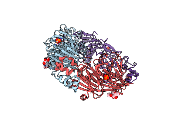
Crystal Structure Of Red Kidney Bean Purple Acid Phosphatase In Complex With Maybridge Fragment Cc24201
Organism: Phaseolus vulgaris
Method: X-RAY DIFFRACTION Resolution:2.30 Å Release Date: 2012-09-19 Classification: HYDROLASE/HYDROLASE INHIBITOR Ligands: ZN, FE, 0LO, GOL, NAG, SO4, EDO, ACT |
 |
Crystal Structure Of Red Kidney Bean Purple Acid Phosphatase In Complex With Maybridge Fragment Cc27209
Organism: Phaseolus vulgaris
Method: X-RAY DIFFRACTION Resolution:2.70 Å Release Date: 2012-09-19 Classification: HYDROLASE/HYDROLASE INHIBITOR Ligands: ZN, FE, GOL, 0LV, SO4, ACT, EDO, NAG |
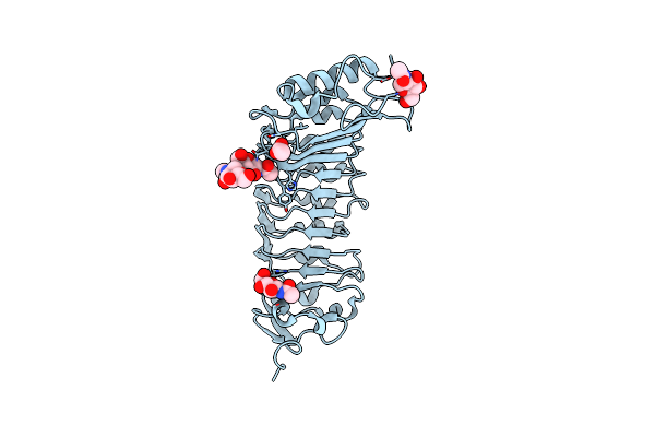 |
The Crystal Structure Of Pgip (Polygalacturonase Inhibiting Protein), A Leucine Rich Repeat Protein Involved In Plant Defense
Organism: Phaseolus vulgaris
Method: X-RAY DIFFRACTION Resolution:1.70 Å Release Date: 2003-07-24 Classification: INHIBITOR Ligands: NAG, ACT |
 |
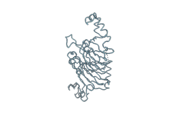
The Structure Of Phaseolin At 2.2 Angstroms Resolution: Implications For A Common Vicilin(Slash)Legumin Structure And The Genetic Engineering Of Seed Storage Proteins
Organism: Phaseolus vulgaris
Method: X-RAY DIFFRACTION Resolution:2.20 Å Release Date: 1994-09-30 Classification: PLANT SEED STORAGE PROTEIN(VICILIN) Ligands: NAG, PO4 |
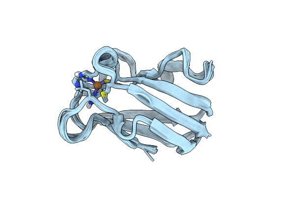 |
High-Resolution Solution Structure Of Reduced French Bean Plastocyanin And Comparison With The Crystal Structure Of Poplar Plastocyanin
Organism: Phaseolus vulgaris
Method: SOLUTION NMR Release Date: 1993-10-31 Classification: ELECTRON TRANSPORT Ligands: CU |
 |
The Three-Dimensional Structure Of The Seed Storage Protein Phaseolin At 3 Angstroms Resolution
Organism: Phaseolus vulgaris
Method: X-RAY DIFFRACTION Resolution:3.00 Å Release Date: 1990-10-15 Classification: PLANT SEED STORAGE PROTEIN (VICILIN) |