
Search Count: 7
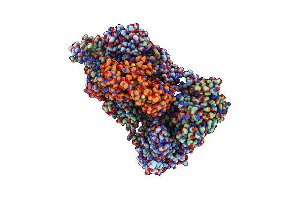 |
Structure Of The Visual Signaling Complex Between Transducin And Phosphodiesterase 6
Organism: Bos taurus
Method: ELECTRON MICROSCOPY Release Date: 2020-10-21 Classification: SIGNALING PROTEIN Ligands: ZN, MG, VDN, 35G, GTP |
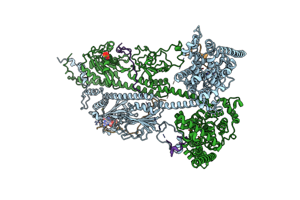 |
Organism: Bos taurus
Method: ELECTRON MICROSCOPY Release Date: 2019-03-06 Classification: SIGNALING PROTEIN Ligands: ZN, MG, 35G |
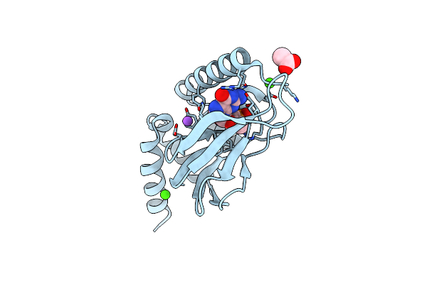 |
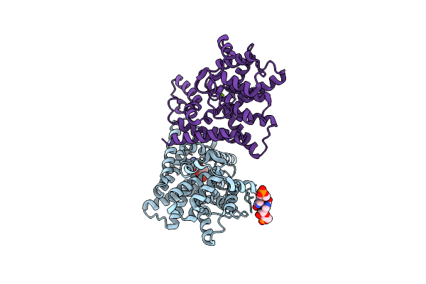
Pkg Ii'S Carboxyl Terminal Cyclic Nucleotide Binding Domain (Cnb-B) In A Complex With Cgmp
Organism: Homo sapiens
Method: X-RAY DIFFRACTION Resolution:1.94 Å Release Date: 2016-01-20 Classification: TRANSFERASE Ligands: 35G, CA, ACT, NA |
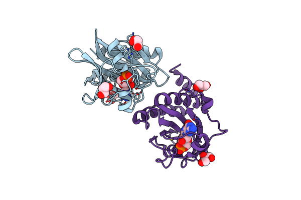 |
Crystal Structure Of The Cgmp-Bound Gaf A Domain From The Photoreceptor Phosphodiesterase 6C
Organism: Gallus gallus
Method: X-RAY DIFFRACTION Resolution:2.57 Å Release Date: 2008-07-01 Classification: HYDROLASE Ligands: 35G, GOL |
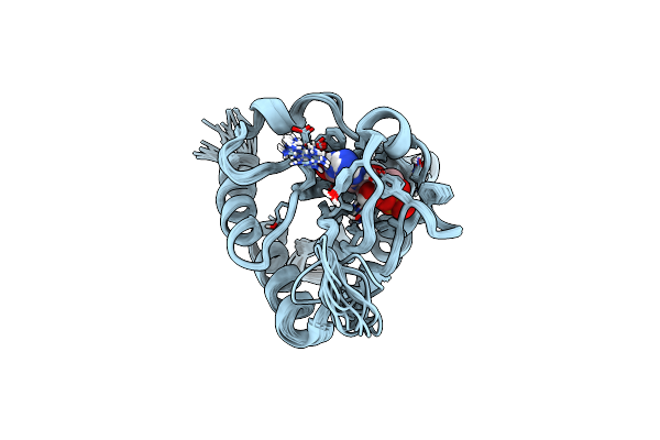 |
Organism: Mus musculus
Method: SOLUTION NMR Release Date: 2008-06-03 Classification: HYDROLASE Ligands: 35G |
 |
Organism: Homo sapiens
Method: X-RAY DIFFRACTION Resolution:1.52 Å Release Date: 2007-03-20 Classification: HYDROLASE Ligands: MG, 35G |
 |
Organism: Bos taurus
Method: X-RAY DIFFRACTION Resolution:2.40 Å Release Date: 1999-02-16 Classification: PHOSPHOTRANSFERASE Ligands: 35G, GDP |