Planned Maintenance: Some services may turn out to be unavailable from 15th January, 2026 to 16th January, 2026. We apologize for the inconvenience!

Planned Maintenance: Some services may turn out to be unavailable from 15th January, 2026 to 16th January, 2026. We apologize for the inconvenience!
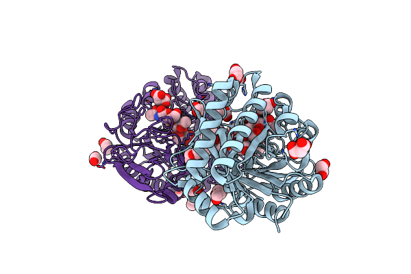 |
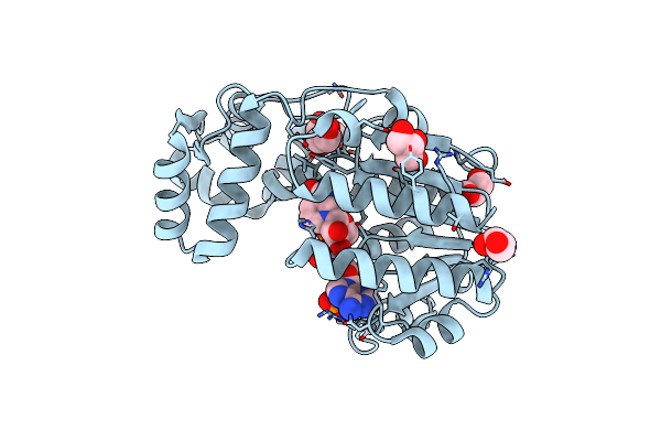
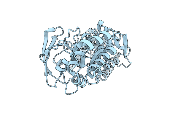
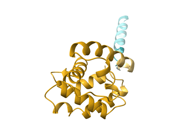
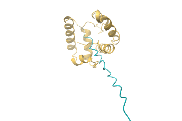
Crystal Structure Of The Catalytic Domain Of Chitiniphilus Shinanonensis Chitinase Chil (Cschil) Complexed With N,N'-Diacetylchitobiose
Organism: Chitiniphilus shinanonensis
Method: X-RAY DIFFRACTION Resolution:1.35 Å Release Date: 2020-09-16 Classification: HYDROLASE Ligands: ACT, MXE, MLI, PGE, EDO, NAG, PG4 |
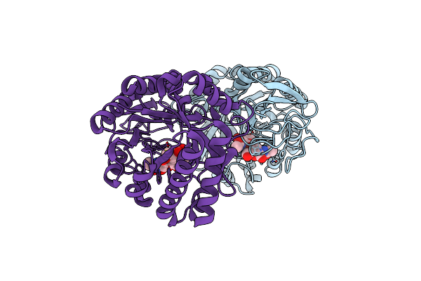 |
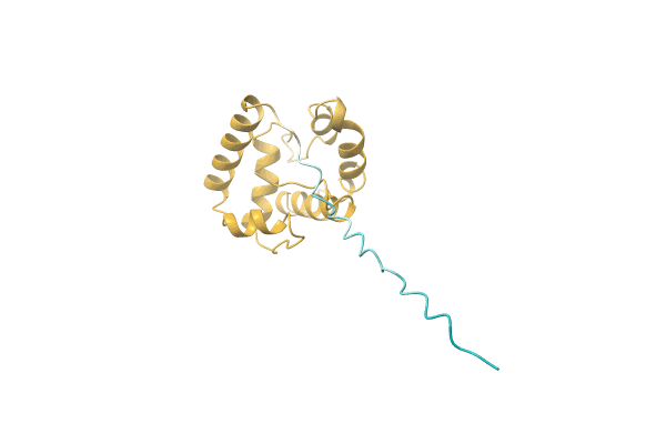
Crystal Structure Of D157N Mutant Of Chitiniphilus Shinanonensis Chitinase Chil (Cschil) Complexed With N,N'-Diacetylchitobiose
Organism: Chitiniphilus shinanonensis
Method: X-RAY DIFFRACTION Resolution:1.95 Å Release Date: 2020-09-16 Classification: HYDROLASE |
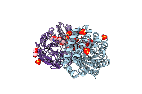 |
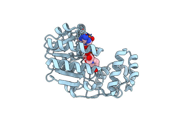
Crystal Structure Of W50A Mutant Of Chitiniphilus Shinanonensis Chitinase Chil (Cschil) Complexed With N,N'-Diacetylchitobiose
Organism: Chitiniphilus shinanonensis
Method: X-RAY DIFFRACTION Resolution:1.50 Å Release Date: 2020-09-16 Classification: HYDROLASE Ligands: SO4, PEG, GOL, EDO |
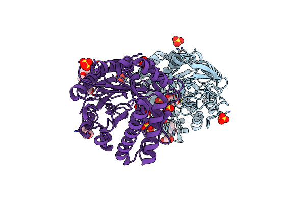 |
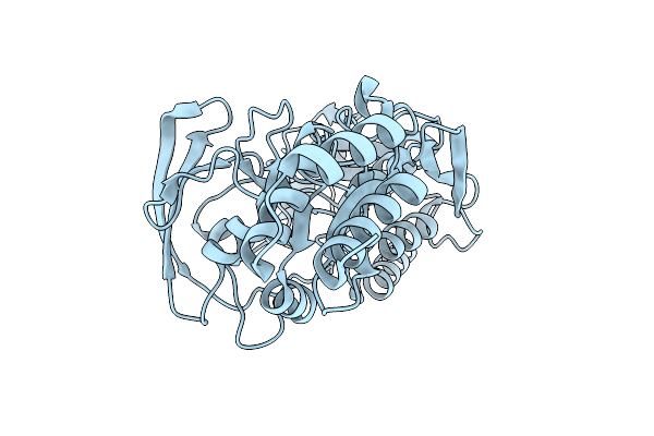
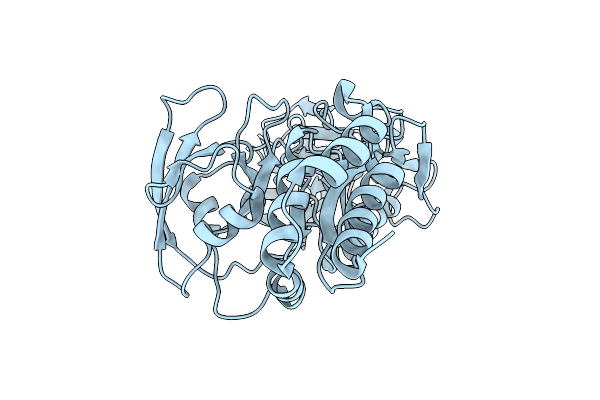
Crystal Structure Of The Catalytic Domain Of Chitinase Chil From Chitiniphilus Shinanonensis (Cschil)
Organism: Chitiniphilus shinanonensis
Method: X-RAY DIFFRACTION Resolution:1.25 Å Release Date: 2020-08-26 Classification: HYDROLASE Ligands: GOL, SO4, EDO, MXE, CL |
 |
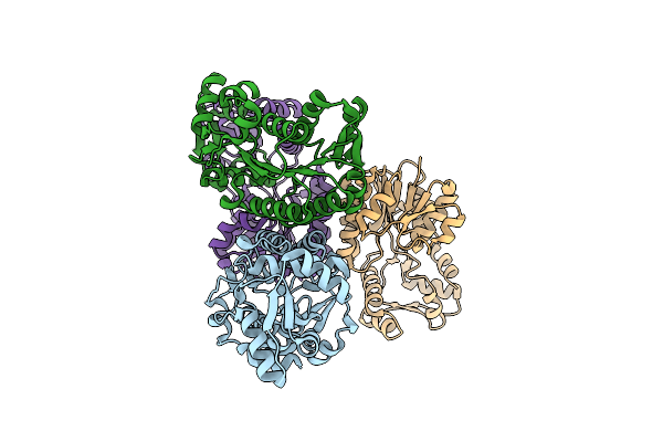
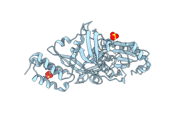
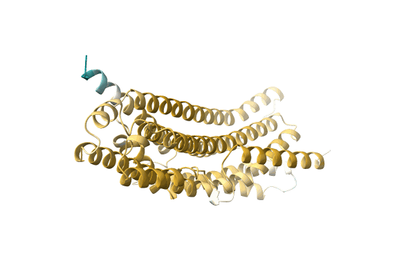
Structural Insight Into Bioremediation Of Triphenylmethane Dyes By Citrobacter Sp. Triphenylmethane Reductase
Organism: Citrobacter sp. my-5
Method: X-RAY DIFFRACTION Resolution:1.96 Å Release Date: 2008-09-23 Classification: OXIDOREDUCTASE Ligands: NAP, GOL |
 |
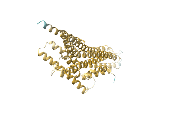
Crystal Structure Of The Citrobacter Sp. Triphenylmethane Reductase Complexed With Nadp(H)
Organism: Citrobacter sp. my-5
Method: X-RAY DIFFRACTION Resolution:2.00 Å Release Date: 2008-09-23 Classification: OXIDOREDUCTASE Ligands: NAP |
 |
Crystal Structure Of The Citrobacter Sp. Triphenylmethane Reductase Complexed With Nadp(H)
Organism: Citrobacter sp. my-5
Method: X-RAY DIFFRACTION Resolution:2.50 Å Release Date: 2008-09-23 Classification: OXIDOREDUCTASE |
 |
Organism: Scenedesmus obliquus
Method: X-RAY DIFFRACTION Resolution:1.80 Å Release Date: 2001-01-18 Classification: HYDROLASE |
 |
Organism: Scenedesmus obliquus
Method: X-RAY DIFFRACTION Resolution:2.00 Å Release Date: 2001-01-18 Classification: HYDROLASE |
 |
Organism: Scenedesmus obliquus
Method: X-RAY DIFFRACTION Resolution:1.90 Å Release Date: 2001-01-18 Classification: HYDROLASE |
 |
Organism: Scenedesmus obliquus
Method: X-RAY DIFFRACTION Resolution:2.10 Å Release Date: 2001-01-18 Classification: HYDROLASE Ligands: SO4 |
 |
 |
 |
 |
 |
 |
 |
 |
 |