
Search Count: 1,594
 |
Organism: Caloramator australicus rc3, Unidentified
Method: X-RAY DIFFRACTION Release Date: 2025-12-10 Classification: METAL BINDING PROTEIN Ligands: ZN, NA, EPE, MG |
 |
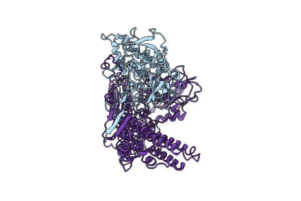
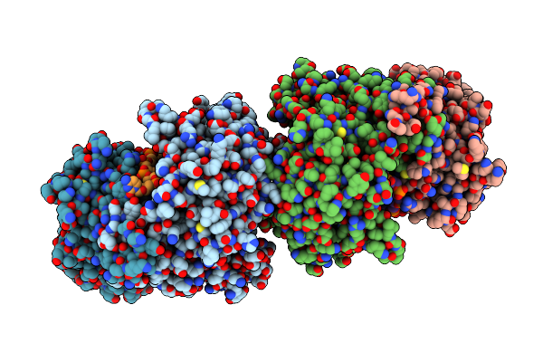
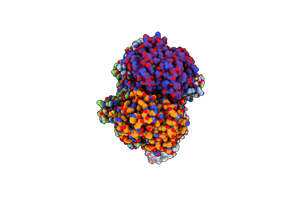
Crystal Structure Of The Catalytic Domain From The Bont-Like Toxin Complex Pg1 Of Paeniclostridium Ghonii
Organism: Paraclostridium ghonii
Method: X-RAY DIFFRACTION Release Date: 2025-10-15 Classification: TOXIN Ligands: ZN, SO4, ACT, GOL |
 |
Crystal Structure Of Dihydrofolate Reductase (Dhfr) From The Filarial Nematode W. Bancrofti In Complex With Nadph And [2-({4-[(2-Amino-4-Oxo-4,7-Dihydro-3H-Pyrrolo[2,3-D]Pyrimidin-5-Yl)Methyl]Benzene-1-Carbonyl}Amino)-4-Methoxyphenyl]Acetic Acid (Tsd10 Or Oed)
Organism: Wuchereria bancrofti
Method: X-RAY DIFFRACTION Release Date: 2025-08-27 Classification: OXIDOREDUCTASE Ligands: NDP, OED |
 |
Crystal Structure Of Dihydrofolate Reductase (Dhfr) From The Filarial Nematode W. Bancrofti In Complex With Nadph And [2-({4-[(2-Amino-4-Oxo-4,7-Dihydro-3H-Pyrrolo[2,3-D]Pyrimidin-5-Yl)Methyl]Benzene-1-Carbonyl}Amino)-4-Cyanophenyl]Acetic Acid (Tsd25 Or Ofd)
Organism: Wuchereria bancrofti
Method: X-RAY DIFFRACTION Release Date: 2025-08-27 Classification: OXIDOREDUCTASE Ligands: NAP, OFD |
 |
Crystal Structure Of Dihydrofolate Reductase (Dhfr) From The Filarial Nematode W. Bancrofti In Complex With Nadph And Methotrexate (Mtx)
Organism: Wuchereria bancrofti
Method: X-RAY DIFFRACTION Release Date: 2025-08-27 Classification: OXIDOREDUCTASE Ligands: NAP, MTX |
 |
Crystal Structure Of Dihydrofolate Reductase (Dhfr) From The Filarial Nematode W. Bancrofti In Complex With Nadph And Antifolate 2-({4-[(2-Amino-4-Oxo-4,7-Dihydro-1H-Pyrrolo[2,3-D]Pyrimidin-5-Yl)Methyl]Benzene-1-Carbonyl}Amino)Benzoic Acid (Og7 Or Tsd001)
Organism: Wuchereria bancrofti
Method: X-RAY DIFFRACTION Release Date: 2025-08-27 Classification: OXIDOREDUCTASE Ligands: NAP, OG7 |
 |
Organism: Flavobacterium sp. 316
Method: X-RAY DIFFRACTION Resolution:1.88 Å Release Date: 2025-04-23 Classification: HYDROLASE |
 |
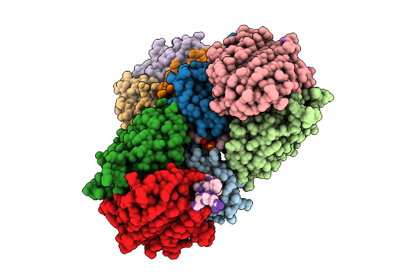
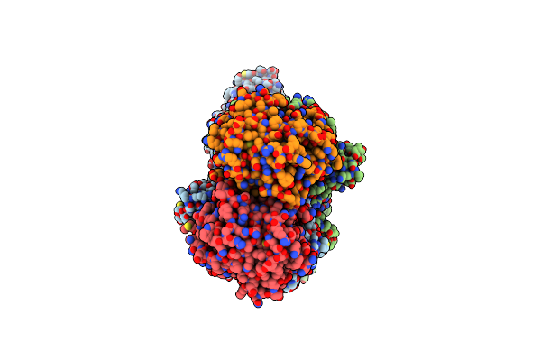
Cryo-Em Structure And Molecular Assembly Of The Bont-Like Toxin Pg1 Complex From Paeniclostridium Ghonii
Organism: Paraclostridium ghonii
Method: ELECTRON MICROSCOPY Resolution:3.20 Å Release Date: 2025-03-26 Classification: TOXIN |
 |
Cryoem Structure Of F-Ena Fibers On The Spores Of Bacillus Thuringiensis Serovar Kurstaki
Organism: Bacillus thuringiensis serovar kurstaki
Method: ELECTRON MICROSCOPY Release Date: 2025-03-12 Classification: PROTEIN FIBRIL |
 |
Organism: Bacillus thuringiensis serovar kurstaki
Method: ELECTRON MICROSCOPY Release Date: 2025-03-12 Classification: PROTEIN FIBRIL |
 |
Organism: Streptantibioticus cattleyicolor
Method: X-RAY DIFFRACTION Release Date: 2025-03-05 Classification: LIGASE Ligands: ADE |
 |
Organism: Maribacter polysiphoniae
Method: ELECTRON MICROSCOPY Release Date: 2024-12-11 Classification: RNA BINDING PROTEIN/DNA/RNA |
 |
Organism: Maribacter polysiphoniae
Method: ELECTRON MICROSCOPY Release Date: 2024-09-11 Classification: DNA BINDING PROTEIN/DNA/RNA |
 |
Organism: Bacillus subtilis, Bacillus phage spbc2
Method: ELECTRON MICROSCOPY Release Date: 2024-08-14 Classification: IMMUNE SYSTEM |
 |
Organism: Bacillus subtilis, Bacillus phage spbeta
Method: ELECTRON MICROSCOPY Release Date: 2024-08-14 Classification: IMMUNE SYSTEM |
 |
Organism: Bacillus subtilis, Bacillus phage spbc2
Method: ELECTRON MICROSCOPY Release Date: 2024-04-10 Classification: ANTIVIRAL PROTEIN |
 |
Organism: Bacillus subtilis, Bacillus phage spbc2
Method: ELECTRON MICROSCOPY Release Date: 2024-04-10 Classification: ANTIVIRAL PROTEIN |
 |
Organism: Bacillus subtilis, Bacillus phage spbc2
Method: ELECTRON MICROSCOPY Release Date: 2024-04-10 Classification: ANTIVIRAL PROTEIN |
 |
Organism: Bacillus subtilis, Bacillus phage spbc2
Method: ELECTRON MICROSCOPY Release Date: 2024-04-10 Classification: ANTIVIRAL PROTEIN Ligands: NAD |
 |
Organism: Brevundimonas diminuta
Method: X-RAY DIFFRACTION Resolution:1.50 Å Release Date: 2024-04-03 Classification: HYDROLASE Ligands: MPD, CL |