
Search Count: 285
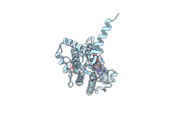 |
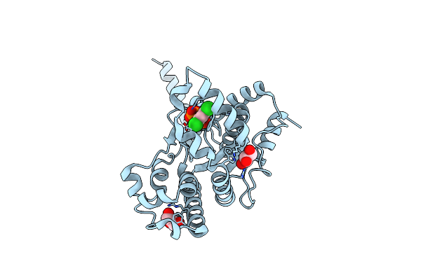
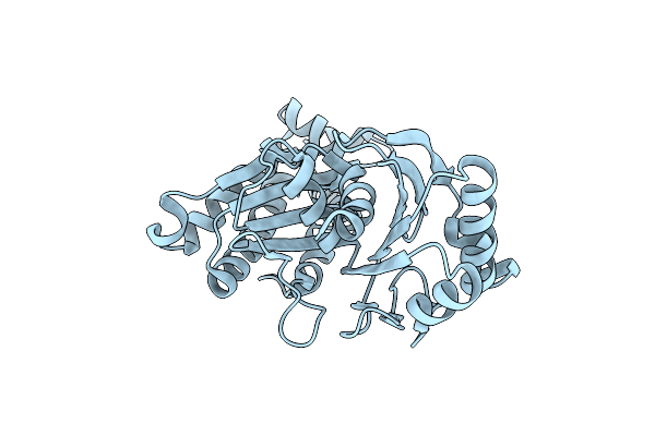
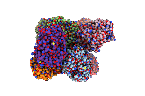
Crystal Structure Of Ppk2 Class Iii In Complex With Adp From Cytophaga Hutchinsonii Atcc 33406
Organism: Cytophaga hutchinsonii (strain atcc 33406 / ncimb 9469)
Method: X-RAY DIFFRACTION Resolution:1.89 Å Release Date: 2019-01-16 Classification: TRANSFERASE Ligands: ADP, GOL |
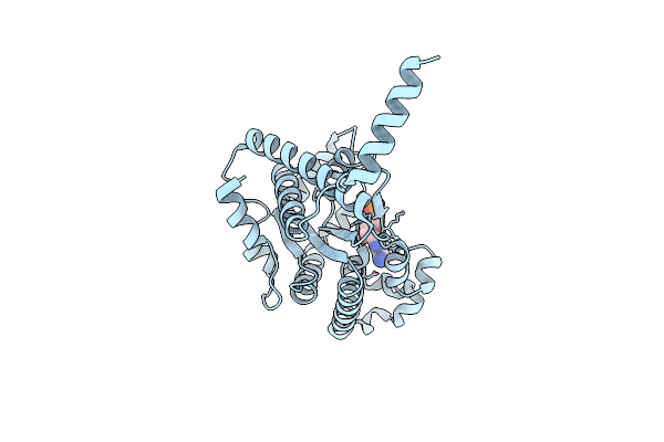 |
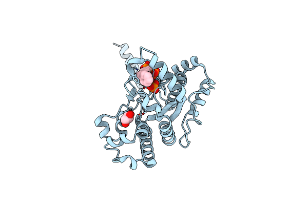
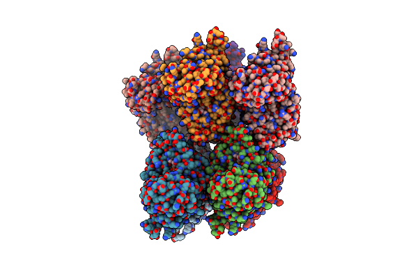
Crystal Structure Of Ppk2 Class Iii In The Complex With Amp From Cytophaga Hutchinsonii Atcc 33406
Organism: Cytophaga hutchinsonii (strain atcc 33406 / ncimb 9469)
Method: X-RAY DIFFRACTION Resolution:2.45 Å Release Date: 2019-01-16 Classification: TRANSFERASE Ligands: AMP, CL |
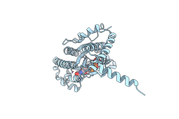 |
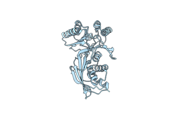
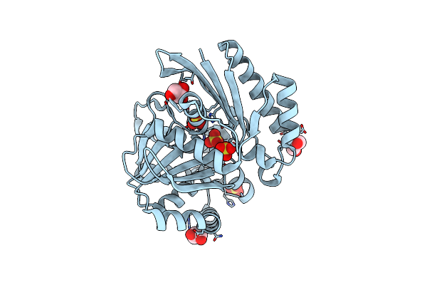
Crystal Structure Of Ppk2 Class Iii In Complex With Guanosine 5-Tetraphosphate
Organism: Cytophaga hutchinsonii (strain atcc 33406 / ncimb 9469)
Method: X-RAY DIFFRACTION Resolution:2.65 Å Release Date: 2019-01-16 Classification: TRANSFERASE Ligands: BKP |
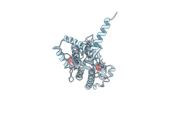 |
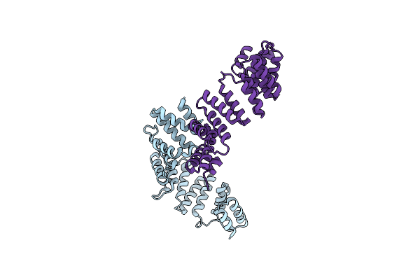
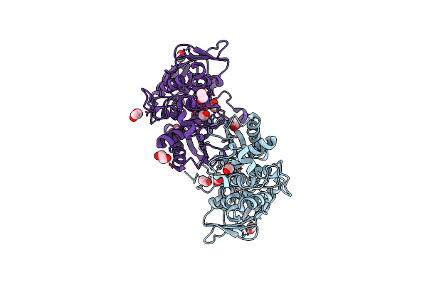
Organism: Cytophaga hutchinsonii (strain atcc 33406 / ncimb 9469)
Method: X-RAY DIFFRACTION Resolution:2.20 Å Release Date: 2019-01-16 Classification: transferase/transferase inhibitor Ligands: BOY, GOL, SRT |
 |
Crystal Structure Of Ppk2 (Class Iii) In Complex With Bisphosphonate Inhibitor (2-((3,5-Dichlorophenyl)Amino)Ethane-1,1-Diyl)Diphosphonic Acid
Organism: Cytophaga hutchinsonii (strain atcc 33406 / ncimb 9469)
Method: X-RAY DIFFRACTION Resolution:2.10 Å Release Date: 2019-01-16 Classification: transferase/transferase inhibitor Ligands: BWJ, GOL |
 |
Organism: Cytophaga hutchinsonii
Method: X-RAY DIFFRACTION Resolution:2.30 Å Release Date: 2019-01-16 Classification: transferase/transferase inhibitor Ligands: C8A, PO4, GOL |
 |
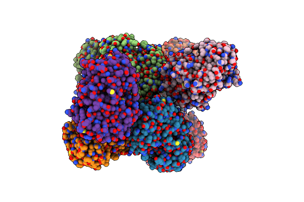
Crystal Structure Of S-Adenosyl-L-Homocysteine Hydrolase From Cytophaga Hutchinsonii In Complex With Adenosine
Organism: Cytophaga hutchinsonii (strain atcc 33406 / ncimb 9469)
Method: X-RAY DIFFRACTION Resolution:2.09 Å Release Date: 2018-08-08 Classification: HYDROLASE Ligands: ADN, NAD, NA, PG4, PEG |
 |
Organism: Cytophaga hutchinsonii (strain atcc 33406 / ncimb 9469)
Method: X-RAY DIFFRACTION Resolution:1.10 Å Release Date: 2016-06-22 Classification: HYDROLASE |
 |
Crystal Structure Of Homoserine Kinase From Cytophaga Hutchinsonii Atcc 33406, Nysgrc Target 032717.
Organism: Cytophaga hutchinsonii
Method: X-RAY DIFFRACTION Resolution:2.60 Å Release Date: 2014-04-02 Classification: TRANSFERASE |
 |
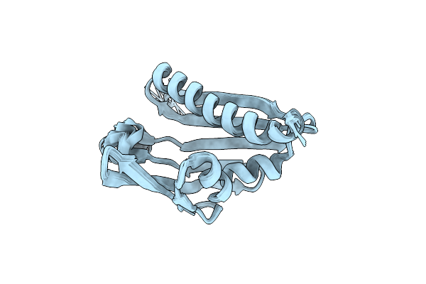
Crystal Structure Of The C-Terminal Part Of The Tpr Repeat-Containing Protein Q11Ti6_Cyth3 From Cytophaga Hutchinsonii. Northeast Structural Genomics Consortium Target Chr11B.
Organism: Cytophaga hutchinsonii
Method: X-RAY DIFFRACTION Resolution:2.28 Å Release Date: 2011-10-19 Classification: Structural Genomics, Unknown Function |
 |
Chemical Shift Assignment And Solution Structure Of Chr145 From Cytophaga Hutchinsonii, Northeast Structural Genomics Consortium Target Chr145
Organism: Cytophaga hutchinsonii
Method: SOLUTION NMR Release Date: 2011-08-17 Classification: STRUCTURAL GENOMICS, UNKNOWN FUNCTION |
 |
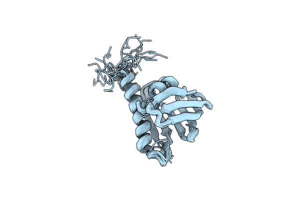
Solution Nmr Structure Of The Ahsa1-Like Protein Chu_1110 From Cytophaga Hutchinsonii, Northeast Structural Genomics Consortium Target Chr152
Organism: Cytophaga hutchinsonii
Method: SOLUTION NMR Release Date: 2011-07-13 Classification: STRUCTURAL GENOMICS, UNKNOWN FUNCTION |
 |
Crystal Structure Of A Putative Sugar Kinase (Chu_1875) From Cytophaga Hutchinsonii Atcc 33406 At 1.65 A Resolution
Organism: Cytophaga hutchinsonii
Method: X-RAY DIFFRACTION Resolution:1.65 Å Release Date: 2011-05-18 Classification: TRANSFERASE |
 |
Solution Structure Of Chr148 From Cytophaga Hutchinsonii, Northeast Structural Genomics Consortium Target Chr148
Organism: Cytophaga hutchinsonii atcc 33406
Method: SOLUTION NMR Release Date: 2011-03-02 Classification: STRUCTURAL GENOMICS, UNKNOWN FUNCTION |
 |
Crystal Structure Of Dipeptide Epimerase From Cytophaga Hutchinsonii Complexed With Mg And Dipeptide D-Ala-L-Val
Organism: Cytophaga hutchinsonii
Method: X-RAY DIFFRACTION Resolution:3.00 Å Release Date: 2011-02-16 Classification: ISOMERASE Ligands: MG, DAL, VAL |
 |
Crystal Structure Of Dipeptide Epimerase From Cytophaga Hutchinsonii Complexed With Mg And Dipeptide D-Ala-L-Ala
Organism: Cytophaga hutchinsonii
Method: X-RAY DIFFRACTION Resolution:3.00 Å Release Date: 2011-02-16 Classification: ISOMERASE Ligands: MG, DAL, ALA |
 |
Putative Antitoxin Component, Chu_2935 Protein, From Xre Family From Prevotella Buccae.
Organism: Cytophaga hutchinsonii
Method: X-RAY DIFFRACTION Resolution:1.65 Å Release Date: 2010-09-08 Classification: structural genomics, unknown function Ligands: PO4 |
 |
Crystal Structure Of Flavoprotein/Dehydrogenase From Cytophaga Hutchinsonii. Northeast Structural Genomics Consortium Target Chr43.
Organism: Cytophaga hutchinsonii
Method: X-RAY DIFFRACTION Resolution:2.60 Å Release Date: 2010-06-30 Classification: OXIDOREDUCTASE Ligands: FAD |
 |
Crystal Structure Of An Exopolyphosphatase (Chu_0316) From Cytophaga Hutchinsonii Atcc 33406 At 1.50 A Resolution
Organism: Cytophaga hutchinsonii
Method: X-RAY DIFFRACTION Resolution:1.50 Å Release Date: 2010-05-12 Classification: HYDROLASE Ligands: CL, SO4, GOL |
 |
Crystal Structure Of Putative Phosphoribosylformylglycinamidine Cyclo-Ligase (Yp_676759.1) From Cytophaga Hutchinsonii Atcc 33406 At 1.50 A Resolution
Organism: Cytophaga hutchinsonii atcc 33406
Method: X-RAY DIFFRACTION Resolution:1.50 Å Release Date: 2009-11-17 Classification: LIGASE Ligands: ACT, EDO |