
Search Count: 3,844
 |
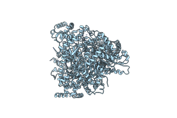
Organism: Micromonospora sp. hm134
Method: X-RAY DIFFRACTION Release Date: 2025-07-16 Classification: BIOSYNTHETIC PROTEIN Ligands: EDO, PEG, CIT, NA |
 |
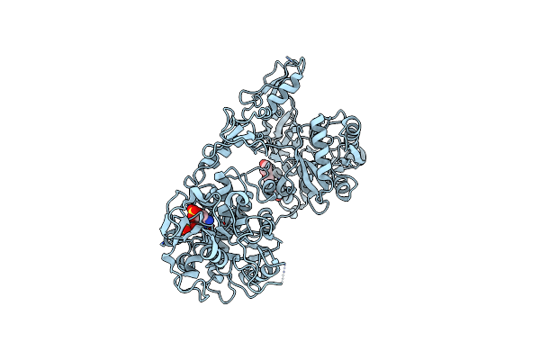
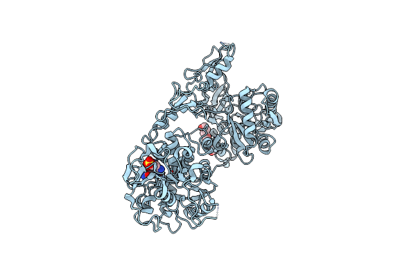
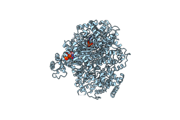
Organism: Pseudorhizobium banfieldiae
Method: X-RAY DIFFRACTION Resolution:1.89 Å Release Date: 2025-05-21 Classification: OXIDOREDUCTASE Ligands: SER, 4MO, F3S, EDO, PEG, MGD, SO4, P33, GOL, O, FES, 1PE |
 |
Organism: Nanorana parkeri
Method: X-RAY DIFFRACTION Resolution:1.80 Å Release Date: 2025-05-07 Classification: ANTITOXIN Ligands: 1PE, YGU |
 |
Organism: Nanorana parkeri
Method: X-RAY DIFFRACTION Resolution:1.90 Å Release Date: 2025-05-07 Classification: ANTITOXIN Ligands: 1PE, YGZ |
 |
Organism: Nanorana parkeri
Method: X-RAY DIFFRACTION Resolution:1.90 Å Release Date: 2025-05-07 Classification: ANTITOXIN Ligands: 1PE, YH9 |
 |
Organism: Nanorana parkeri
Method: X-RAY DIFFRACTION Resolution:1.90 Å Release Date: 2025-05-07 Classification: ANTITOXIN Ligands: 1PE, YGF |
 |
Organism: Nanorana parkeri
Method: X-RAY DIFFRACTION Resolution:1.95 Å Release Date: 2025-05-07 Classification: ANTITOXIN Ligands: 1PE, YGQ |
 |
Organism: Orthotospovirus tomatomaculae
Method: ELECTRON MICROSCOPY Release Date: 2025-04-16 Classification: VIRAL PROTEIN Ligands: RTP |
 |
Organism: Orthotospovirus tomatomaculae
Method: ELECTRON MICROSCOPY Release Date: 2025-01-29 Classification: VIRAL PROTEIN |
 |
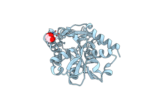
Organism: Nocardioides sp. s-1144
Method: X-RAY DIFFRACTION Resolution:1.55 Å Release Date: 2024-12-04 Classification: HYDROLASE Ligands: SO4 |
 |
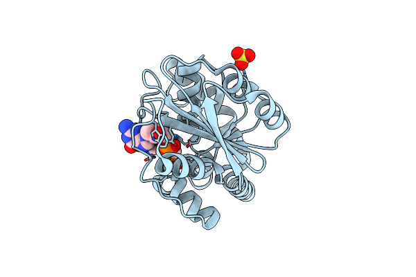
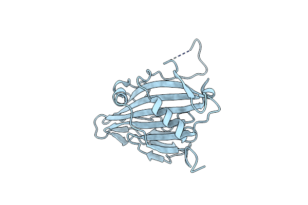
Structure Of A Solute Binding Protein From Desulfonauticus Sp. Bound To L-Tryptophan
Organism: Desulfonauticus sp. 38_4375
Method: X-RAY DIFFRACTION Resolution:1.80 Å Release Date: 2024-11-06 Classification: PEPTIDE BINDING PROTEIN Ligands: TRP, EDO, PEG, ACT, PG4 |
 |
Organism: Orthotospovirus tomatomaculae
Method: ELECTRON MICROSCOPY Release Date: 2024-09-25 Classification: VIRAL PROTEIN Ligands: RBV |
 |
Organism: Orthotospovirus tomatomaculae, Synthetic construct
Method: ELECTRON MICROSCOPY Release Date: 2024-09-25 Classification: VIRAL PROTEIN/RNA |
 |
Organism: Orthotospovirus tomatomaculae
Method: ELECTRON MICROSCOPY Release Date: 2024-09-25 Classification: VIRAL PROTEIN |
 |
Organism: Alcaligenes ammonioxydans
Method: X-RAY DIFFRACTION Resolution:2.10 Å Release Date: 2024-09-04 Classification: TRANSFERASE |
 |
Organism: Streptomyces cavourensis
Method: X-RAY DIFFRACTION Resolution:2.20 Å Release Date: 2024-06-05 Classification: OXIDOREDUCTASE |
 |
Organism: Thermothielavioides terrestris
Method: ELECTRON MICROSCOPY Release Date: 2024-03-13 Classification: PROTEIN TRANSPORT |
 |
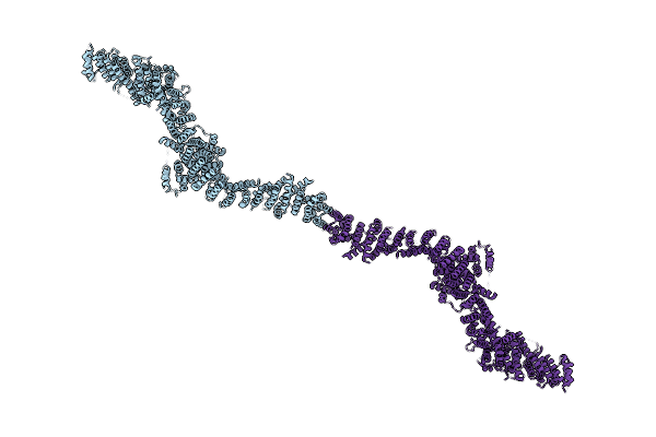
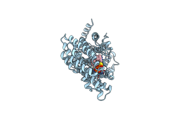
Crystal Structure Of The Histidine Kinase Domain Of Bacteriophytochrome Rpbphp2
Organism: Rhodopseudomonas palustris cga009
Method: X-RAY DIFFRACTION Resolution:3.19 Å Release Date: 2023-11-22 Classification: SIGNALING PROTEIN, TRANSFERASE Ligands: ATP, MG |
 |
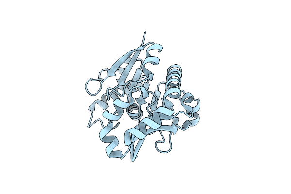
Crystal Structure Of Loop Deletion Aiox Mutant From Pseudorhizobium Sp. Str. Nt-26
Organism: Pseudorhizobium banfieldiae
Method: X-RAY DIFFRACTION Resolution:1.79 Å Release Date: 2023-11-08 Classification: SIGNALING PROTEIN Ligands: GOL |
 |
Organism: Artabotrys hexapetalus
Method: ELECTRON MICROSCOPY Release Date: 2023-10-25 Classification: PLANT PROTEIN Ligands: FPS, MG |