Planned Maintenance: Some services may turn out to be unavailable from 15th January, 2026 to 16th January, 2026. We apologize for the inconvenience!

Planned Maintenance: Some services may turn out to be unavailable from 15th January, 2026 to 16th January, 2026. We apologize for the inconvenience!
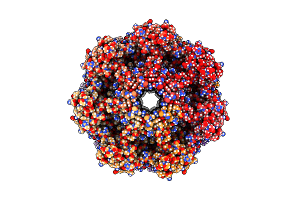 |
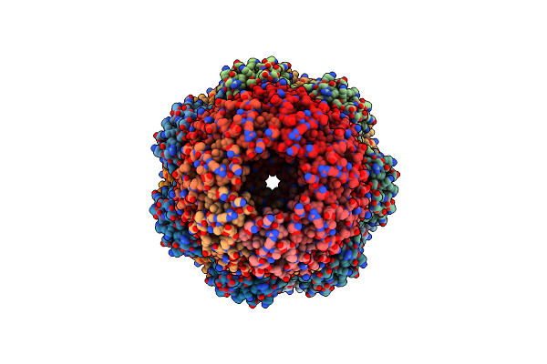
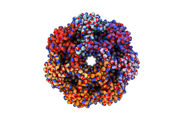
Organism: Thermoplasma acidophilum
Method: ELECTRON MICROSCOPY Release Date: 2024-10-30 Classification: HYDROLASE |
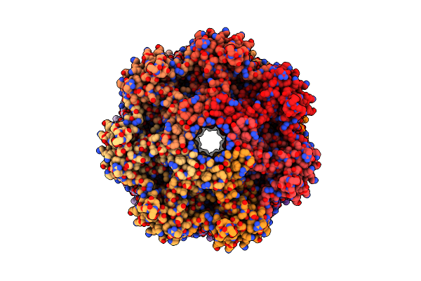 |
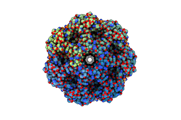
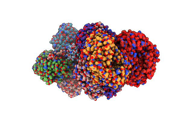
Organism: Thermoplasma acidophilum
Method: ELECTRON MICROSCOPY Release Date: 2023-08-30 Classification: HYDROLASE |
 |
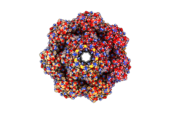
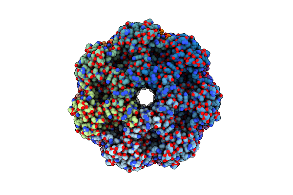
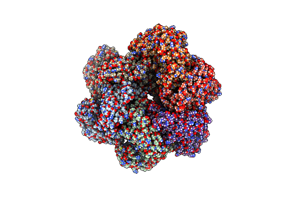
Organism: Thermoplasma acidophilum
Method: ELECTRON MICROSCOPY Release Date: 2023-08-30 Classification: HYDROLASE |
 |
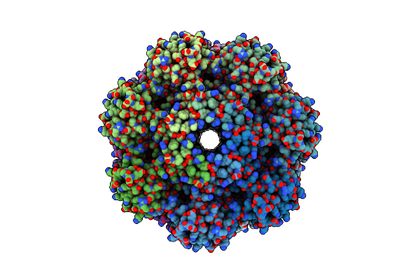
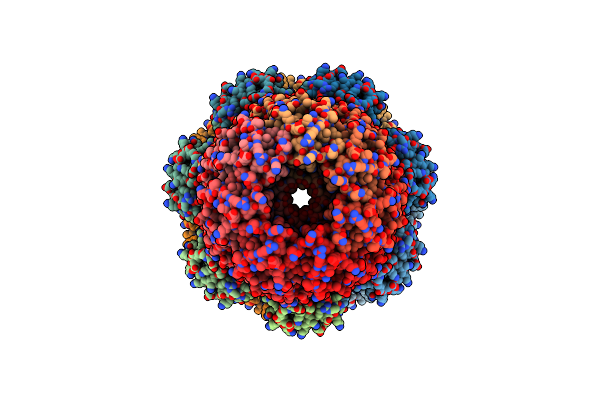
Organism: Thermoplasma acidophilum
Method: ELECTRON MICROSCOPY Release Date: 2023-08-30 Classification: HYDROLASE Ligands: XIB |
 |
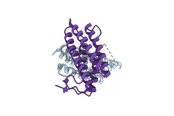
Cryo-Em Imaging Scaffold Subunits A And B Used To Display Kras G12C Complex With Gdp
Organism: Thermoplasma acidophilum, Synthetic construct
Method: ELECTRON MICROSCOPY Release Date: 2023-08-09 Classification: STRUCTURAL PROTEIN |
 |
Organism: Thermoplasma acidophilum
Method: X-RAY DIFFRACTION Resolution:2.10 Å Release Date: 2022-04-06 Classification: LYASE |
 |
Organism: Solimonas sp. k1w22b-7
Method: X-RAY DIFFRACTION Release Date: 2022-01-26 Classification: HYDROLASE |
 |
Allosteric Coupling Between Alpha-Rings Of The 20S Proteasome, Archaea 20S Proteasome Singly Capped With A Pan Complex
Organism: Thermoplasma acidophilum
Method: ELECTRON MICROSCOPY Release Date: 2020-09-09 Classification: HYDROLASE |
 |
Allosteric Coupling Between Alpha-Rings Of The 20S Proteasome, 20S Singly Capped With A Pa26/V230F
Organism: Thermoplasma acidophilum, Trypanosoma brucei
Method: ELECTRON MICROSCOPY Release Date: 2020-09-09 Classification: HYDROLASE |
 |
Allosteric Coupling Between Alpha-Rings Of 20S Proteasome, 20S Proteasome Singly Capped With A Pa26/E102A_Panc, Together With Lfp Incubation
Organism: Thermoplasma acidophilum, Trypanosoma brucei brucei
Method: ELECTRON MICROSCOPY Release Date: 2020-09-09 Classification: HYDROLASE |
 |
Allosteric Coupling Between Alpha-Rings Of 20S Proteasome, 20S Proteasome With Singly Capped Pan Complex
Organism: Thermoplasma acidophilum
Method: ELECTRON MICROSCOPY Release Date: 2020-09-09 Classification: HYDROLASE |
 |
Allosteric Couple Between Alpha Rings Of The 20S Proteasome. 20S Proteasome Singly Capped By Pa26/E102A, C-Terminus Replaced By Pan C-Terminus
Organism: Thermoplasma acidophilum, Trypanosoma brucei brucei
Method: ELECTRON MICROSCOPY Release Date: 2020-09-09 Classification: HYDROLASE |
 |
Accidental Minimum Contact Crystal Lattice Formed By A Redesigned Protein Oligomer
Organism: Thermoplasma acidophilum
Method: X-RAY DIFFRACTION Resolution:4.10 Å Release Date: 2018-05-23 Classification: DE NOVO PROTEIN |
 |
2.8 A Resolution Reconstruction Of The Thermoplasma Acidophilum 20S Proteasome Using Cryo-Electron Microscopy
Organism: Thermoplasma acidophilum
Method: ELECTRON MICROSCOPY Release Date: 2017-12-27 Classification: HYDROLASE |
 |
Organism: Thermoplasma acidophilum
Method: ELECTRON MICROSCOPY Release Date: 2017-06-14 Classification: HYDROLASE |
 |
Organism: Thermoplasma acidophilum
Method: ELECTRON MICROSCOPY Release Date: 2017-06-14 Classification: HYDROLASE |
 |
Wild-Type Glyceraldehyde Dehydrogenase From Thermoplasma Acidophilum In Complex With Nadp
Organism: Thermoplasma acidophilum
Method: X-RAY DIFFRACTION Resolution:2.05 Å Release Date: 2017-04-05 Classification: OXIDOREDUCTASE Ligands: NAP |
 |
Crystal Structure Of The T33-51H Designed Self-Assembling Protein Tetrahedron
Organism: Thermoplasma acidophilum, Pyrococcus horikoshii
Method: X-RAY DIFFRACTION Resolution:3.40 Å Release Date: 2016-08-10 Classification: DE NOVO PROTEIN |
 |
Organism: Thermoplasma acidophilum
Method: ELECTRON MICROSCOPY Resolution:7.00 Å Release Date: 2016-07-27 Classification: HYDROLASE |
 |
Organism: Thermoplasma acidophilum
Method: ELECTRON MICROSCOPY Resolution:7.80 Å Release Date: 2016-07-27 Classification: HYDROLASE |