Planned Maintenance: Some services may turn out to be unavailable from 15th January, 2026 to 16th January, 2026. We apologize for the inconvenience!

Planned Maintenance: Some services may turn out to be unavailable from 15th January, 2026 to 16th January, 2026. We apologize for the inconvenience!
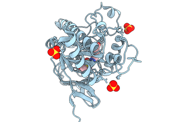 |
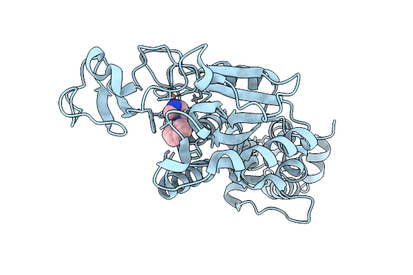
Isopenicillin N Synthase N252E Variant In Complex With Fe And Ipn After O2 Exposure
Organism: Aspergillus nidulans fgsc a4
Method: X-RAY DIFFRACTION Release Date: 2026-01-14 Classification: OXIDOREDUCTASE Ligands: FE, SO4, IP1 |
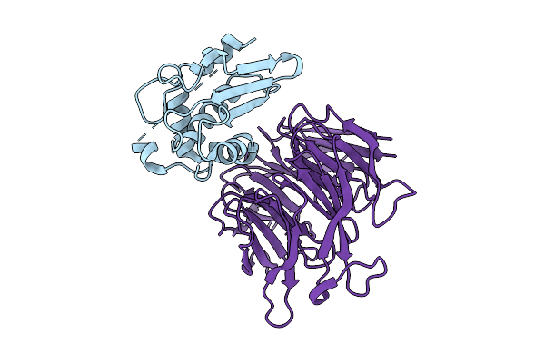 |
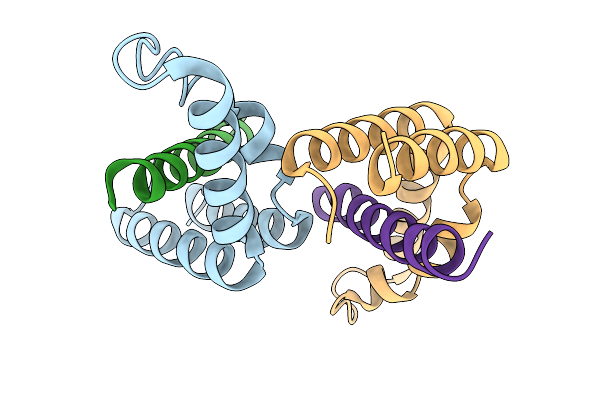
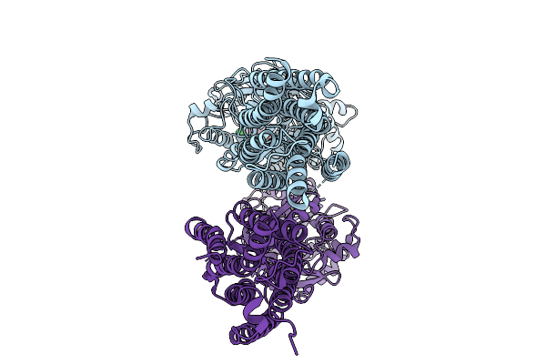
Organism: Drosophila melanogaster
Method: X-RAY DIFFRACTION Release Date: 2025-12-24 Classification: METAL BINDING PROTEIN |
 |
Organism: Azotobacter vinelandii dj
Method: X-RAY DIFFRACTION Release Date: 2025-12-03 Classification: METAL BINDING PROTEIN Ligands: SF4, S5Q, PEG, EDO, 1PE, PGE, MG, PG4 |
 |
Organism: Drosophila melanogaster
Method: ELECTRON MICROSCOPY Release Date: 2025-12-03 Classification: MEMBRANE PROTEIN Ligands: K |
 |
Organism: Drosophila melanogaster, Homo sapiens
Method: ELECTRON MICROSCOPY Release Date: 2025-10-29 Classification: LIGASE Ligands: AMP |
 |
Organism: Drosophila melanogaster, Homo sapiens
Method: ELECTRON MICROSCOPY Release Date: 2025-10-29 Classification: LIGASE Ligands: AMP |
 |
Organism: Drosophila melanogaster, Homo sapiens
Method: ELECTRON MICROSCOPY Release Date: 2025-10-29 Classification: LIGASE Ligands: AMP |
 |
Organism: Drosophila melanogaster, Homo sapiens
Method: ELECTRON MICROSCOPY Release Date: 2025-10-22 Classification: MEMBRANE PROTEIN Ligands: ABU |
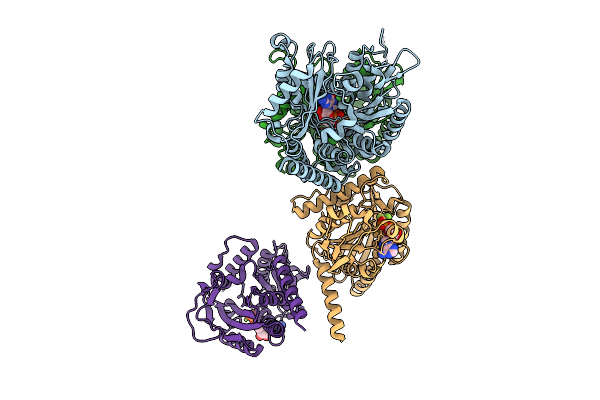 |
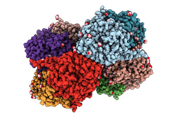
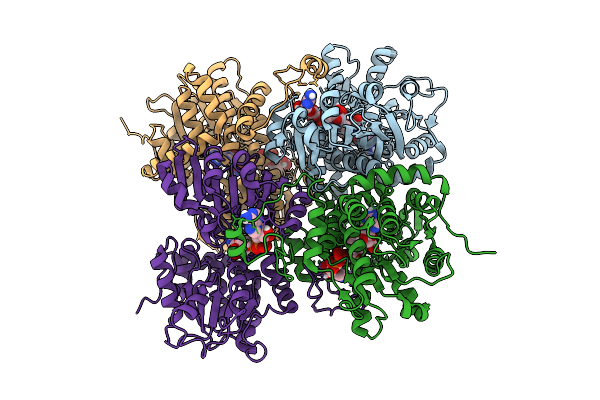
Organism: Drosophila melanogaster, Sus scrofa
Method: ELECTRON MICROSCOPY Release Date: 2025-10-15 Classification: CELL CYCLE Ligands: GTP, GDP, ADP, MG, ALF |
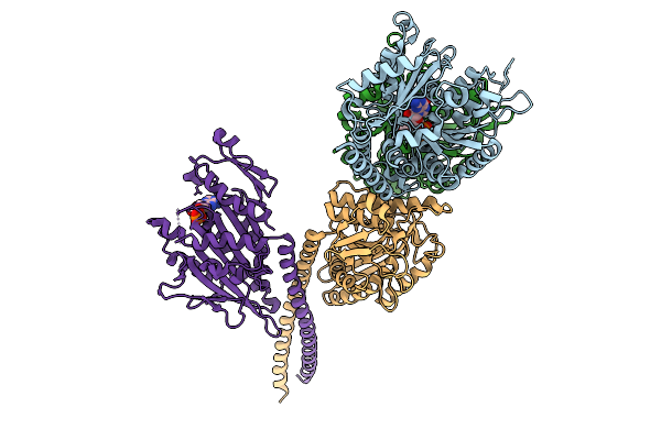 |
Organism: Drosophila melanogaster, Sus scrofa
Method: ELECTRON MICROSCOPY Release Date: 2025-10-08 Classification: CELL CYCLE Ligands: GTP, GDP, MG, ADP |
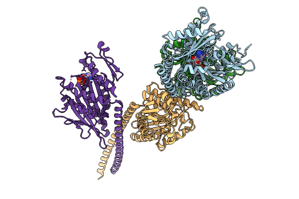 |
Organism: Drosophila melanogaster, Sus scrofa
Method: ELECTRON MICROSCOPY Release Date: 2025-10-08 Classification: CELL CYCLE Ligands: GTP, GDP, MG, ADP |
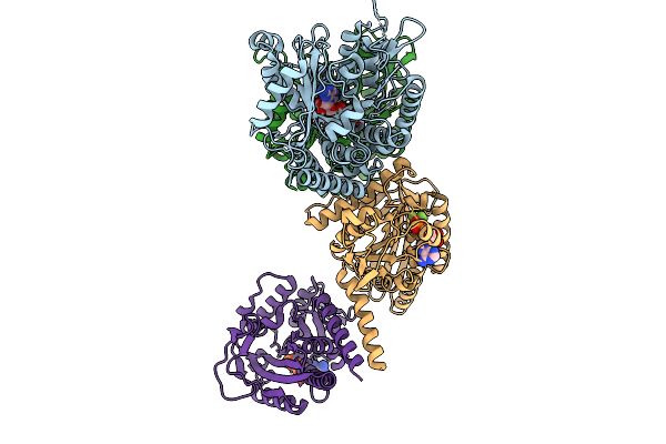 |
Organism: Drosophila melanogaster, Sus scrofa
Method: ELECTRON MICROSCOPY Release Date: 2025-10-08 Classification: CELL CYCLE Ligands: GTP, GDP, ADP, MG, ALF |
 |
Organism: Drosophila melanogaster
Method: X-RAY DIFFRACTION Release Date: 2025-10-08 Classification: TRANSCRIPTION |
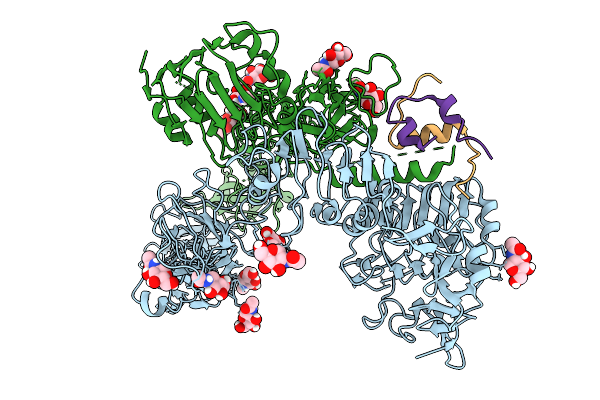 |
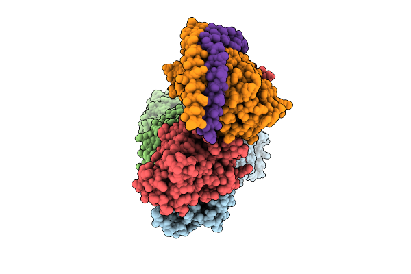
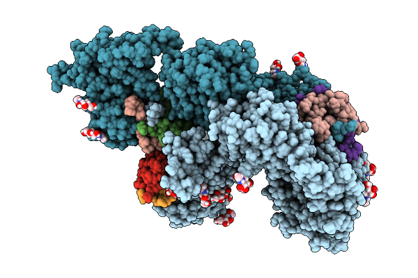
Cryo-Em Structure Of Drosophila Melanogaster Insulin Receptor (Dmir) Bound With One Dilp1, Asymmetric Conformation
Organism: Drosophila melanogaster
Method: ELECTRON MICROSCOPY Release Date: 2025-10-01 Classification: STRUCTURAL PROTEIN Ligands: NAG |
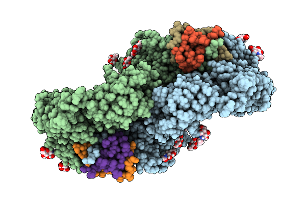 |
Cryo-Em Structure Of Drosophila Melanogaster Insulin Receptor (Dmir) Bound With Two Dilp1, Symmetric Conformation
Organism: Drosophila melanogaster
Method: ELECTRON MICROSCOPY Release Date: 2025-10-01 Classification: STRUCTURAL PROTEIN Ligands: NAG |
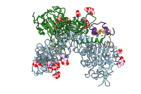 |
Cryo-Em Structure Of Drosophila Melanogaster Insulin Receptor (Dmir) Bound With One Dilp2, Asymmetric Conformation
Organism: Drosophila melanogaster
Method: ELECTRON MICROSCOPY Release Date: 2025-10-01 Classification: STRUCTURAL PROTEIN Ligands: NAG |
 |
Cryo-Em Structure Of Drosophila Melanogaster Insulin Receptor (Dmir) Bound With Three Dilp5, Asymmetric Conformation
Organism: Drosophila melanogaster
Method: ELECTRON MICROSCOPY Release Date: 2025-10-01 Classification: STRUCTURAL PROTEIN Ligands: NAG |
 |
Organism: Drosophila melanogaster
Method: ELECTRON MICROSCOPY Release Date: 2025-10-01 Classification: MEMBRANE PROTEIN Ligands: 2BV |
 |
Organism: Drosophila melanogaster
Method: X-RAY DIFFRACTION Release Date: 2025-09-17 Classification: HYDROLASE Ligands: NAD, ADN |
 |
Organism: Drosophila melanogaster
Method: X-RAY DIFFRACTION Release Date: 2025-09-03 Classification: HYDROLASE Ligands: ZN, A1EOF, FE2 |