
Search Count: 4,468
 |
Organism: Rabbit hemorrhagic disease virus
Method: ELECTRON MICROSCOPY Release Date: 2025-12-03 Classification: VIRAL PROTEIN |
 |
Organism: Rabbit hemorrhagic disease virus
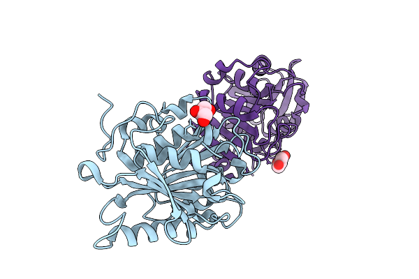
Method: X-RAY DIFFRACTION Release Date: 2025-12-03 Classification: VIRAL PROTEIN Ligands: EDO, NHE, TRS, PEG |
 |
Organism: Rabbit hemorrhagic disease virus
Method: ELECTRON MICROSCOPY Release Date: 2025-11-26 Classification: VIRUS LIKE PARTICLE |
 |
Organism: Rabbit hemorrhagic disease virus
Method: ELECTRON MICROSCOPY Release Date: 2025-11-26 Classification: VIRAL PROTEIN |
 |
Organism: Rabbit hemorrhagic disease virus
Method: ELECTRON MICROSCOPY Release Date: 2025-11-26 Classification: VIRUS |
 |
Organism: Acinetobacter silvestris
Method: X-RAY DIFFRACTION Release Date: 2025-11-26 Classification: CELL ADHESION Ligands: ACT |
 |
Organism: Thermosynechococcus vestitus bp-1
Method: X-RAY DIFFRACTION Release Date: 2025-11-26 Classification: HYDROLASE Ligands: GOL, CA, TLA |
 |
Crystal Structure Of Tegh116 From Thermosynechococcus Elongatus With Glucose
Organism: Thermosynechococcus vestitus bp-1
Method: X-RAY DIFFRACTION Release Date: 2025-11-26 Classification: HYDROLASE Ligands: BGC, GOL, CA, TLA |
 |
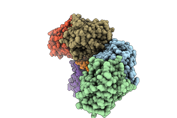
Cryo-Em Structure Of The G Protein-Coupled Receptor 1 (Gpr1) Bound To Chemerin And Beta-Arrestin 1 (Conformation 1)
Organism: Homo sapiens, Escherichia phage ecszw-2
Method: ELECTRON MICROSCOPY Release Date: 2025-11-19 Classification: MEMBRANE PROTEIN/IMMUNE SYSTEM |
 |
Cryo-Em Structure Of The G Protein-Coupled Receptor 1 (Gpr1) Bound To Chemerin And Beta-Arrestin 1 (Conformation 2)
Organism: Homo sapiens, Escherichia phage ecszw-2
Method: ELECTRON MICROSCOPY Release Date: 2025-11-19 Classification: MEMBRANE PROTEIN/IMMUNE SYSTEM |
 |
Cryo-Em Structure Of The G Protein-Coupled Receptor 1 (Gpr1) Bound To Chemerin And Beta-Arrestin 1 (Conformation 3)
Organism: Homo sapiens, Escherichia phage ecszw-2
Method: ELECTRON MICROSCOPY Release Date: 2025-11-19 Classification: MEMBRANE PROTEIN/IMMUNE SYSTEM |
 |
Cryo-Em Structure Of The G Protein-Coupled Receptor 1 (Gpr1) Bound To Chemerin And Beta-Arrestin 1 (Conformation 4)
Organism: Homo sapiens, Escherichia phage ecszw-2
Method: ELECTRON MICROSCOPY Release Date: 2025-11-19 Classification: MEMBRANE PROTEIN/IMMUNE SYSTEM |
 |
Composite Map Of The G Protein-Coupled Receptor 1 (Gpr1) Bound To Chemerin And Beta-Arrestin 2
Organism: Homo sapiens, Escherichia phage ecszw-2
Method: ELECTRON MICROSCOPY Release Date: 2025-11-19 Classification: MEMBRANE PROTEIN/IMMUNE SYSTEM Ligands: Y01 |
 |
Cryo-Em Structure Of The G Protein-Coupled Receptor 1 (Gpr1) Bound To Beta-Arrestin 1 In Ligand-Free State
Organism: Homo sapiens, Escherichia phage ecszw-2
Method: ELECTRON MICROSCOPY Release Date: 2025-11-19 Classification: MEMBRANE PROTEIN/IMMUNE SYSTEM Ligands: PAM |
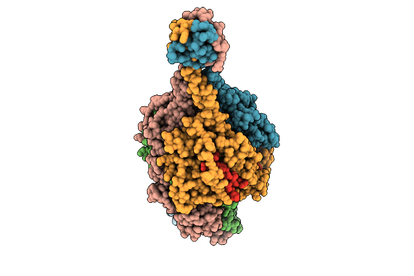 |
Respiratory Syncytial Virus Pre-F Trimer Bound By Neutralizing Antibody Pr306007
Organism: Respiratory syncytial virus a2, Homo sapiens
Method: ELECTRON MICROSCOPY Release Date: 2025-11-12 Classification: STRUCTURAL PROTEIN/IMMUNE SYSTEM |
 |
Organism: Pseudomonadota bacterium
Method: X-RAY DIFFRACTION Release Date: 2025-10-22 Classification: HYDROLASE Ligands: GOL |
 |
Crystal Structure Of The Nitrilase Superfamily Protein Cj1056C From Campylobacter Jejuni In Space Group P212121
Organism: Campylobacter jejuni
Method: X-RAY DIFFRACTION Release Date: 2025-10-08 Classification: HYDROLASE |
 |
Crystal Structure Of The Nitrilase Superfamily Protein Cj1056C From Campylobacter Jejuni In Space Group P21
Organism: Campylobacter jejuni
Method: X-RAY DIFFRACTION Release Date: 2025-10-08 Classification: HYDROLASE |
 |
Organism: Cyclocybe aegerita
Method: X-RAY DIFFRACTION Release Date: 2025-09-24 Classification: OXIDOREDUCTASE Ligands: HEM, MG, NAG |
 |
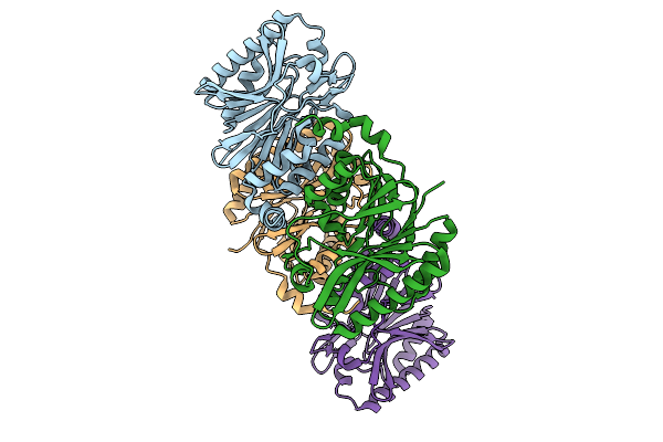
Structural Basis Of The Bifunctionality Of M. Salinexigens Zyf650T Glucosylglycerol Phosphorylase In Glucosylglycerol Catabolism
Organism: Marinobacter salinexigens
Method: X-RAY DIFFRACTION Release Date: 2025-09-10 Classification: STRUCTURAL PROTEIN Ligands: GLC, GOL, NA, G1P |