
Search Count: 326
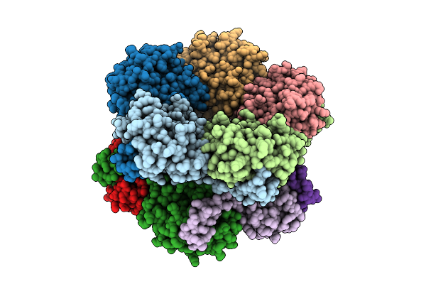 |
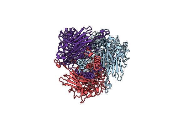
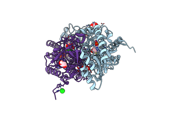
Selective Production Of Versatile L-Glyceraldehyde From C1 And/Or C2 Aldehydes
Organism: Gilliamella apicola
Method: X-RAY DIFFRACTION Resolution:2.00 Å Release Date: 2025-11-12 Classification: SUGAR BINDING PROTEIN |
 |
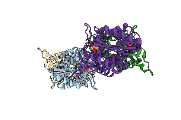
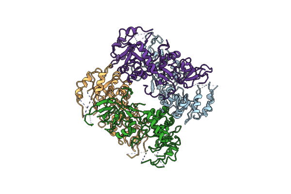
Organism: Aeromonas dhakensis
Method: X-RAY DIFFRACTION Resolution:2.27 Å Release Date: 2025-04-23 Classification: HYDROLASE |
 |
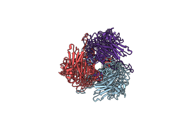
Crystal Structure Of Pfld Bound To 1,5-Anhydromannitol-6-Phosphate In Streptococcus Dysgalactiae Subsp. Equisimilis
Organism: Streptococcus dysgalactiae subsp. equisimilis
Method: X-RAY DIFFRACTION Resolution:2.34 Å Release Date: 2024-10-02 Classification: LYASE Ligands: 7P6 |
 |
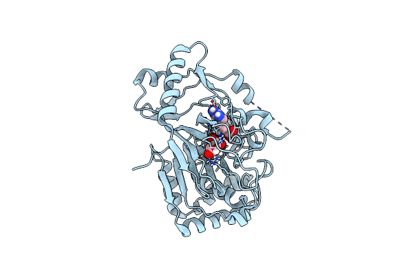
Organism: Streptosporangium roseum, Streptomyces vinaceus
Method: X-RAY DIFFRACTION Resolution:2.00 Å Release Date: 2024-01-10 Classification: TRANSFERASE Ligands: PEG |
 |
Organism: Streptosporangium roseum, Streptomyces vinaceus
Method: X-RAY DIFFRACTION Resolution:2.02 Å Release Date: 2024-01-10 Classification: TRANSFERASE Ligands: PEG, ATP |
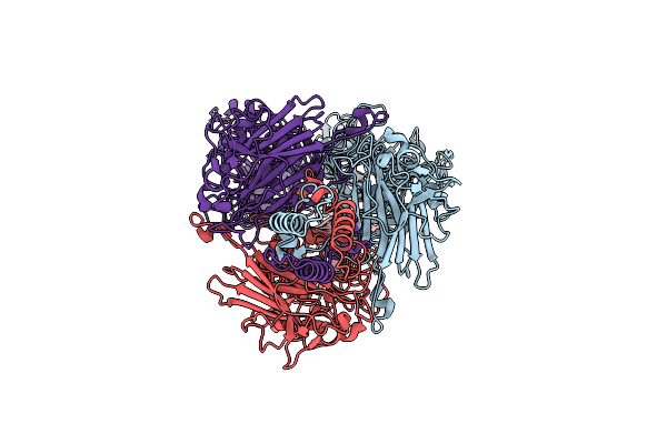 |
Cryo-Em Structure Of Depo32, A Klebsiella Phage Depolymerase Targets The K2 Serotype K. Pneumoniae
Organism: Klebsiella phage gh-k3
Method: ELECTRON MICROSCOPY Resolution:2.32 Å Release Date: 2023-08-30 Classification: LYASE |
 |
Cryo-Em Structure Of Depo32, A Klebsiella Phage Depolymerase Targets The K2 Serotype K. Pneumoniae
Organism: Klebsiella phage gh-k3
Method: ELECTRON MICROSCOPY Resolution:2.46 Å Release Date: 2023-08-30 Classification: HYDROLASE |
 |
Organism: Geobacillus sp. 12amor1, Bacillus oceanisediminis 2691
Method: X-RAY DIFFRACTION Resolution:1.85 Å Release Date: 2022-03-16 Classification: HYDROLASE Ligands: GOL |
 |
Organism: Klebsiella phage gh-k3
Method: ELECTRON MICROSCOPY Release Date: 2021-08-25 Classification: SUGAR BINDING PROTEIN |
 |
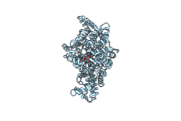
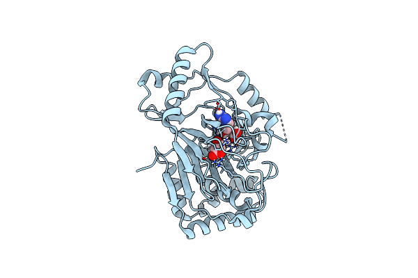
Arginine Hydroxylase Vioc In Complex With Arg, 2Og And Fe Under Anaerobic Environment Using Ft-Ssx Methods
Organism: Streptomyces vinaceus
Method: X-RAY DIFFRACTION Resolution:1.86 Å Release Date: 2020-09-09 Classification: OXIDOREDUCTASE Ligands: ARG, AKG, FE |
 |
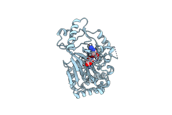
Arginine Hydroxylase Vioc In Complex With (3S)-Oh-Arg, Succinate And Fe After Oxygen Exposure Using Ft-Ssx Methods
Organism: Streptomyces vinaceus
Method: X-RAY DIFFRACTION Resolution:1.70 Å Release Date: 2020-09-09 Classification: OXIDOREDUCTASE Ligands: SIN, FE, ZZU |
 |
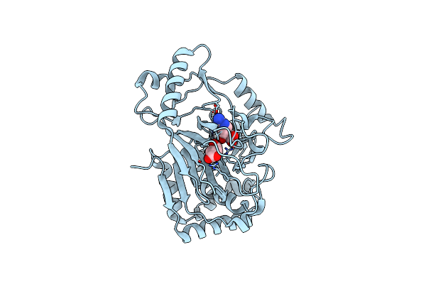
Vioc L-Arginine Hydroxylase Bound To Fe(Ii), L-Arginine, And 2-Oxo-Glutaric Acid
Organism: Streptomyces vinaceus
Method: X-RAY DIFFRACTION Resolution:1.60 Å Release Date: 2017-09-06 Classification: OXIDOREDUCTASE Ligands: FE2, AKG, ARG |
 |
Vioc L-Arginine Hydroxylase Bound To Fe(Ii), 3S-Hydroxy-L-Arginine, And 2Og
Organism: Streptomyces vinaceus
Method: X-RAY DIFFRACTION Resolution:1.80 Å Release Date: 2017-09-06 Classification: OXIDOREDUCTASE Ligands: AKG, FE2, ZZU |
 |
Vioc L-Arginine Hydroxylase Bound To Fe(Ii), L-Arginine, And A Peroxysuccinate Intermediate
Organism: Streptomyces vinaceus
Method: X-RAY DIFFRACTION Resolution:1.79 Å Release Date: 2017-09-06 Classification: OXIDOREDUCTASE Ligands: FE2, ARG, OKG |
 |
Vioc L-Arginine Hydroxylase Bound To Fe(Ii), 3S-Hydroxy-L-Arginine, And Succinate
Organism: Streptomyces vinaceus
Method: X-RAY DIFFRACTION Resolution:1.99 Å Release Date: 2017-09-06 Classification: OXIDOREDUCTASE Ligands: FE2, SIN, ZZU |
 |
Organism: Streptomyces vinaceus
Method: X-RAY DIFFRACTION Resolution:1.67 Å Release Date: 2017-09-06 Classification: OXIDOREDUCTASE Ligands: FE2, SIN, ARG |
 |
Vioc L-Arginine Hydroxylase Bound To The Vanadyl Ion, L-Arginine, And Succinate
Organism: Streptomyces vinaceus
Method: X-RAY DIFFRACTION Resolution:1.55 Å Release Date: 2017-09-06 Classification: OXIDOREDUCTASE Ligands: SIN, ARG, VVO |
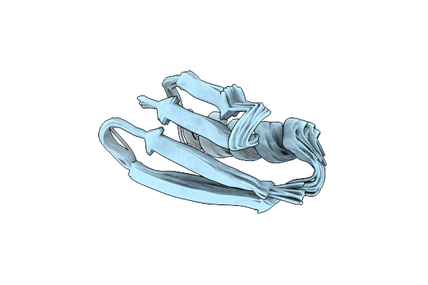 |
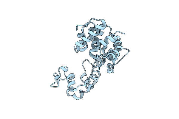
Solid-State Mas Nmr Structure Of Immunoglobulin Beta 1 Binding Domain Of Protein G (Gb1)
Organism: Streptococcus dysgalactiae subsp. equisimilis
Method: SOLID-STATE NMR Release Date: 2016-08-10 Classification: IMMUNE SYSTEM |
 |
Organism: Aeromonas hydrophila subsp. hydrophila
Method: X-RAY DIFFRACTION Resolution:1.45 Å Release Date: 2014-01-15 Classification: ISOMERASE Ligands: PLP, CL, GOL, NI |
 |
Organism: Aeromonas hydrophila subsp. hydrophila
Method: X-RAY DIFFRACTION Resolution:3.25 Å Release Date: 2014-01-15 Classification: ISOMERASE |