
Search Count: 130
 |
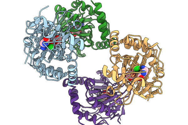
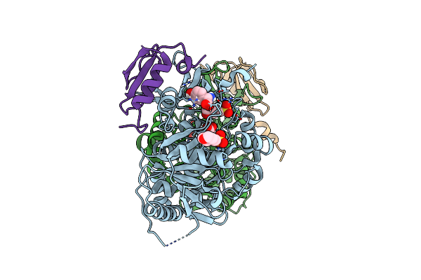
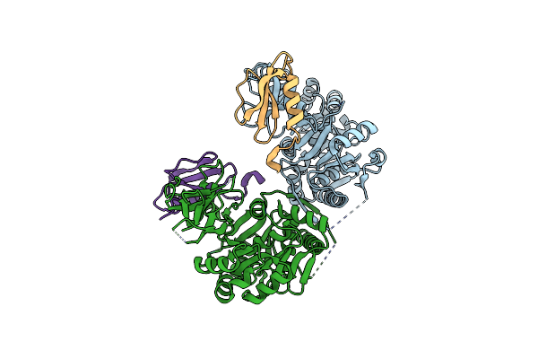
Organism: Actinoplanes teichomyceticus
Method: X-RAY DIFFRACTION Release Date: 2025-11-19 Classification: BIOSYNTHETIC PROTEIN Ligands: LYS, AKG, FE2, CL, SIN, YL0, A1CA1 |
 |
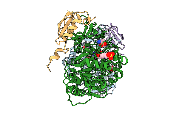
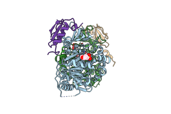
Organism: Actinoplanes teichomyceticus
Method: X-RAY DIFFRACTION Release Date: 2025-11-19 Classification: BIOSYNTHETIC PROTEIN Ligands: LYS, SIN, PGE, VVO, CL, FLC, V |
 |
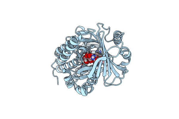
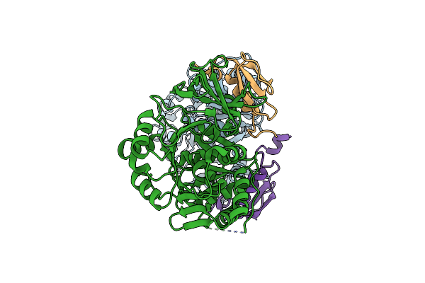
Organism: Actinoplanes teichomyceticus
Method: X-RAY DIFFRACTION Release Date: 2025-11-19 Classification: BIOSYNTHETIC PROTEIN Ligands: LYS, SIN, VVO, CL |
 |
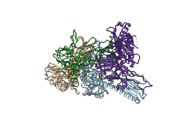
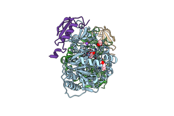
Organism: Actinoplanes teichomyceticus
Method: X-RAY DIFFRACTION Release Date: 2025-11-19 Classification: BIOSYNTHETIC PROTEIN Ligands: LYS, SIN, VVO, CL, V |
 |
Organism: Actinoplanes teichomyceticus
Method: X-RAY DIFFRACTION Release Date: 2025-11-19 Classification: BIOSYNTHETIC PROTEIN Ligands: LYS, SIN, VVO, CL, V |
 |
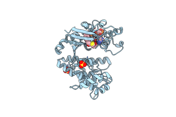
A1 Tei + D-Hpg: Adenylation Domain 1 Core Construct From Teicoplanin Biosynthesis With D-4-Hydroxyphenylglycine
Organism: Actinoplanes teichomyceticus
Method: X-RAY DIFFRACTION Release Date: 2025-08-13 Classification: LIGASE Ligands: GHP, MES, BO3 |
 |
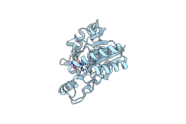
Organism: Legionella clemsonensis
Method: X-RAY DIFFRACTION Resolution:1.80 Å Release Date: 2024-07-03 Classification: OXIDOREDUCTASE Ligands: FAD |
 |
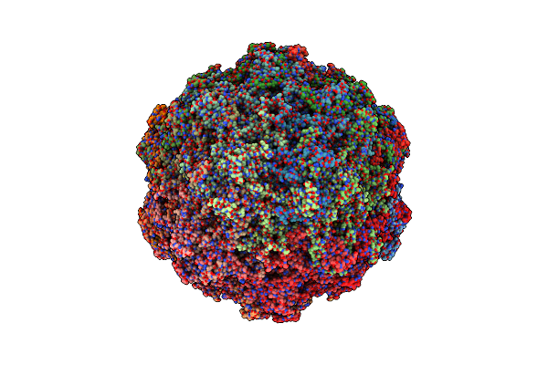
Organism: Lederbergvirus
Method: ELECTRON MICROSCOPY Release Date: 2024-01-31 Classification: VIRUS LIKE PARTICLE |
 |
A1 Tei + Hpg: Adenylation Domain 1 Core Construct From Teicoplanin Biosynthesis; 4-Hydroxyphenylglycine Bound
Organism: Actinoplanes teichomyceticus
Method: X-RAY DIFFRACTION Resolution:1.64 Å Release Date: 2023-12-06 Classification: LIGASE Ligands: MES, D4P |
 |
Organism: Actinoplanes teichomyceticus
Method: X-RAY DIFFRACTION Resolution:1.81 Å Release Date: 2023-12-06 Classification: LIGASE Ligands: GOL, MES |
 |
A1 Int Graft: Adenylation Domain 1 Core Construct From Teicoplanin Biosynthesis, Intermediate Selection Pocket Graft
Organism: Actinoplanes teichomyceticus
Method: X-RAY DIFFRACTION Resolution:2.70 Å Release Date: 2023-12-06 Classification: LIGASE |
 |
A1 Leu Graft + Leu: Adenylation Domain 1 Core Construct From Teicoplanin Biosynthesis, Leucine Selection Pocket Graft; Leucine Bound
Organism: Actinoplanes teichomyceticus
Method: X-RAY DIFFRACTION Resolution:1.89 Å Release Date: 2023-12-06 Classification: LIGASE Ligands: LEU, PEG, ACY, NA |
 |
A1 Ancala: Adenylation Domain 1 Core Construct From Ancestral Reconstruction Of Glycopeptide Antibiotic Biosynthesis, Alanine Selection Pocket
Organism: Actinoplanes, Actinoplanes teichomyceticus
Method: X-RAY DIFFRACTION Resolution:3.12 Å Release Date: 2023-12-06 Classification: LIGASE |
 |
The Crystal Structure Of The Complement Inhibitory Domain Of Borrelia Hermsii Fbpc.
Organism: Borrelia hermsii hs1
Method: X-RAY DIFFRACTION Resolution:1.50 Å Release Date: 2023-06-07 Classification: IMMUNE SYSTEM Ligands: MG |
 |
 |
The Crystal Structure Of Acyltransferase Mutant, Orf11*-W163A, In Complex With Octanoyl-Coa
Organism: Actinoplanes teichomyceticus
Method: X-RAY DIFFRACTION Resolution:1.73 Å Release Date: 2019-10-09 Classification: TRANSFERASE Ligands: CO8, SO4 |
 |
Crystal Structure Of Deacetylase Triple Mutant (Orf2*T) That Involving In Teicoplanin Biosynthetic Pathway
Organism: Actinoplanes teichomyceticus
Method: X-RAY DIFFRACTION Resolution:1.29 Å Release Date: 2019-10-09 Classification: OXIDOREDUCTASE Ligands: IMD, CO |
 |
Organism: Actinoplanes teichomyceticus
Method: X-RAY DIFFRACTION Resolution:1.52 Å Release Date: 2019-09-11 Classification: TRANSFERASE Ligands: IMD, ACT |
 |
X-Ray Crystal Structure Of Pf-M1 In Complex With Inhibitor (6Da) And Catalytic Zinc Ion
Organism: Plasmodium falciparum (isolate fcb1 / columbia)
Method: X-RAY DIFFRACTION Resolution:1.82 Å Release Date: 2018-12-26 Classification: hydrolase/hydrolase inhibitor Ligands: ZN, J0Y, GOL, MG |
 |
X-Ray Crystal Structure Of Pf-M1 In Complex With Inhibitor (6H) And Catalytic Zinc Ion
Organism: Plasmodium falciparum (isolate fcb1 / columbia)
Method: X-RAY DIFFRACTION Resolution:1.35 Å Release Date: 2018-12-26 Classification: hydrolase/hydrolase inhibitor Ligands: ZN, J1G, DMS, GOL, MG |