
Search Count: 55
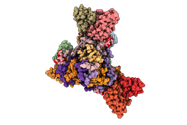 |
A Broadly-Neutralizing Antibody Against Ebolavirus Glycoprotein That Can Potentiate The Breadth And Neutralization Potency Of Other Anti-Glycoprotein Antibodies
Organism: Oryctolagus cuniculus, Ebolavirus
Method: ELECTRON MICROSCOPY Release Date: 2025-11-05 Classification: VIRAL PROTEIN/IMMUNE SYSTEM Ligands: NAG |
 |
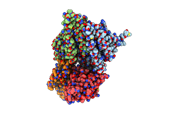
Organism: Homo sapiens, Sudan ebolavirus
Method: ELECTRON MICROSCOPY Release Date: 2025-02-05 Classification: VIRAL PROTEIN |
 |
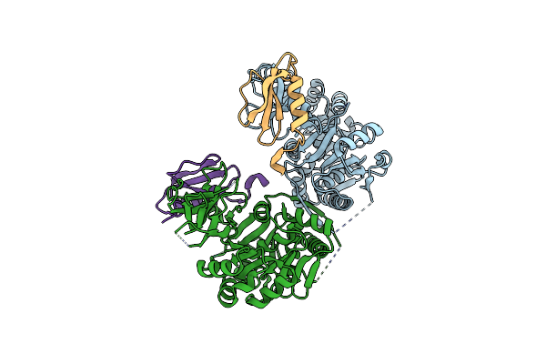
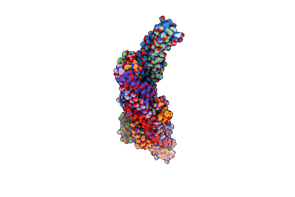
Organism: Zaire ebolavirus, Homo sapiens
Method: ELECTRON MICROSCOPY Release Date: 2025-01-29 Classification: VIRAL PROTEIN/RNA |
 |
A1 Ancala: Adenylation Domain 1 Core Construct From Ancestral Reconstruction Of Glycopeptide Antibiotic Biosynthesis, Alanine Selection Pocket
Organism: Actinoplanes, Actinoplanes teichomyceticus
Method: X-RAY DIFFRACTION Resolution:3.12 Å Release Date: 2023-12-06 Classification: LIGASE |
 |
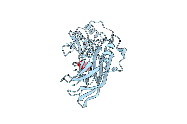
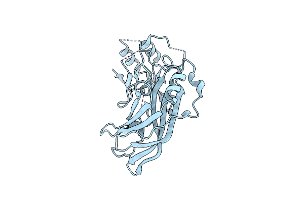
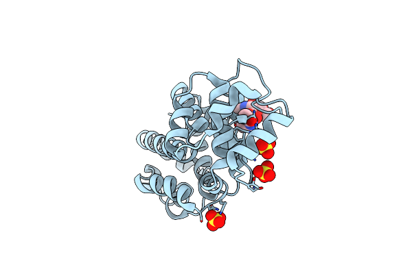
Organism: Sudan ebolavirus
Method: X-RAY DIFFRACTION Resolution:1.60 Å Release Date: 2023-09-27 Classification: VIRAL PROTEIN Ligands: SAL |
 |
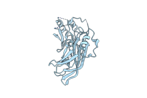
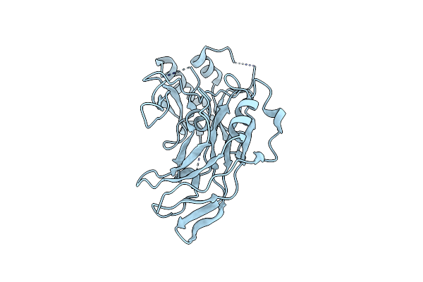
Organism: Sudan ebolavirus
Method: X-RAY DIFFRACTION Resolution:1.80 Å Release Date: 2023-09-27 Classification: VIRAL PROTEIN Ligands: SAL |
 |
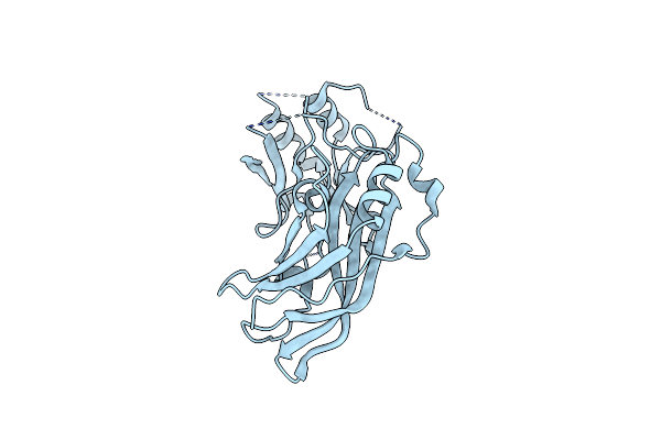
Organism: Sudan ebolavirus
Method: X-RAY DIFFRACTION Resolution:1.90 Å Release Date: 2023-09-13 Classification: VIRAL PROTEIN |
 |
Organism: Sudan ebolavirus
Method: X-RAY DIFFRACTION Resolution:2.00 Å Release Date: 2023-09-13 Classification: VIRAL PROTEIN |
 |
Organism: Sudan ebolavirus
Method: X-RAY DIFFRACTION Resolution:1.75 Å Release Date: 2023-06-21 Classification: VIRAL PROTEIN |
 |
Organism: Sudan ebolavirus
Method: X-RAY DIFFRACTION Resolution:1.70 Å Release Date: 2023-06-21 Classification: VIRAL PROTEIN |
 |
Organism: Sudan ebolavirus
Method: X-RAY DIFFRACTION Resolution:1.53 Å Release Date: 2023-06-21 Classification: VIRAL PROTEIN |
 |
Hypothetical Protein Uy81_C0065G0003 Residues 18-54 From Candidatus Giovannonibacteria Bacterium Fused To Gcn4 Adaptors
Organism: Candidatus giovannonibacteria bacterium gw2011_gwa2_53_7
Method: X-RAY DIFFRACTION Resolution:2.70 Å Release Date: 2022-05-04 Classification: PROTEIN FIBRIL |
 |
Structure Of Ebov Gp Lacking The Mucin-Like Domain With 1C11 Scfv And 1C3 Fab Bound
Organism: Ebola virus - gabon (1994-1997), Zaire ebolavirus, Homo sapiens
Method: ELECTRON MICROSCOPY Release Date: 2022-04-06 Classification: VIRAL PROTEIN/Immune System |
 |
Organism: Ebola virus, Homo sapiens, Zaire ebolavirus
Method: ELECTRON MICROSCOPY Release Date: 2021-06-23 Classification: VIRAL PROTEIN/Immune System Ligands: NAG |
 |
Organism: Zaire ebolavirus
Method: X-RAY DIFFRACTION Resolution:3.50 Å Release Date: 2020-04-15 Classification: VIRAL PROTEIN Ligands: NAG |
 |
Organism: Ebola virus, Homo sapiens, Zaire ebolavirus
Method: ELECTRON MICROSCOPY Release Date: 2020-03-11 Classification: immune system, viral protein Ligands: NAG |
 |
Organism: Oropouche virus
Method: X-RAY DIFFRACTION Resolution:2.09 Å Release Date: 2019-02-27 Classification: VIRAL PROTEIN Ligands: SO4, CL |
 |
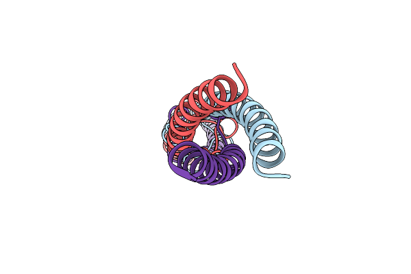
Crystal Structure Of The Oligomerization Domain Of Vp35 From Ebola Virus, Mercury Derivative
Organism: Zaire ebolavirus
Method: X-RAY DIFFRACTION Resolution:3.49 Å Release Date: 2018-10-10 Classification: VIRAL PROTEIN Ligands: HG |
 |
Organism: Reston ebolavirus
Method: X-RAY DIFFRACTION Resolution:2.43 Å Release Date: 2018-10-10 Classification: VIRAL PROTEIN |
 |
Crystal Structure Of The Oligomerization Domain Of Vp35 From Reston Virus, Mercury Derivative
Organism: Reston ebolavirus
Method: X-RAY DIFFRACTION Resolution:3.15 Å Release Date: 2018-10-10 Classification: VIRAL PROTEIN Ligands: MBO |