
Search Count: 794
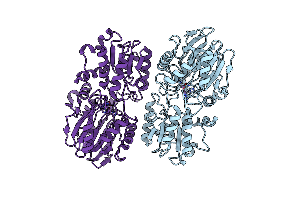 |
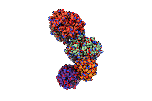
Organism: Staphylococcus epidermidis atcc 12228
Method: X-RAY DIFFRACTION Resolution:2.99 Å Release Date: 2020-10-07 Classification: METAL BINDING PROTEIN Ligands: MN |
 |
Organism: Staphylococcus epidermidis atcc 12228
Method: X-RAY DIFFRACTION Resolution:2.70 Å Release Date: 2020-10-07 Classification: METAL BINDING PROTEIN Ligands: MN |
 |
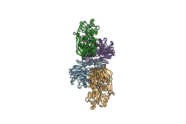
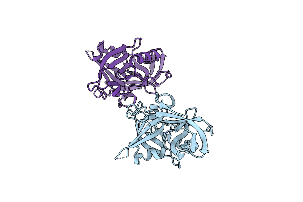
Structure Of N-Terminus Locked Esp With Eight Pro-Peptide Residues - V67C, D255C
Organism: Staphylococcus epidermidis (strain atcc 12228)
Method: X-RAY DIFFRACTION Resolution:2.08 Å Release Date: 2019-09-04 Classification: HYDROLASE |
 |
Structure Of N-Terminus Locked Esp With One Pro-Peptide Residue - V67C, D255C
Organism: Staphylococcus epidermidis (strain atcc 12228)
Method: X-RAY DIFFRACTION Resolution:2.07 Å Release Date: 2019-08-28 Classification: HYDROLASE |
 |
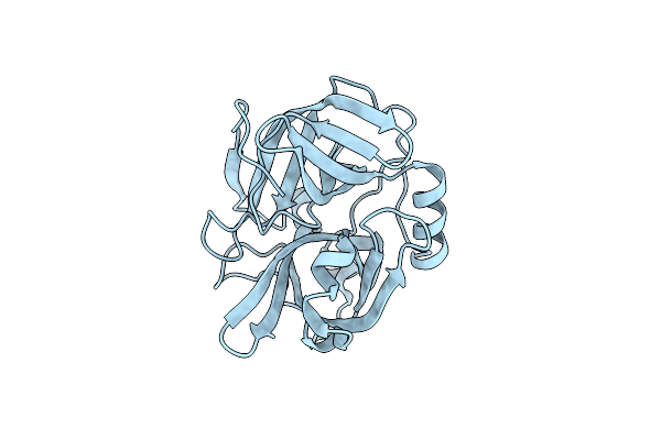
Organism: Staphylococcus epidermidis (strain atcc 12228)
Method: X-RAY DIFFRACTION Resolution:1.85 Å Release Date: 2019-08-21 Classification: HYDROLASE |
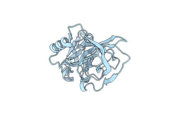 |
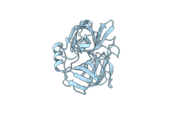
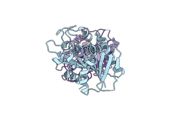
Structure Of Active-Site Serine Mutant Of Esp, Serine Protease From Staphylococcus Epidermidis
Organism: Staphylococcus epidermidis (strain atcc 12228)
Method: X-RAY DIFFRACTION Resolution:1.20 Å Release Date: 2019-08-14 Classification: HYDROLASE |
 |
Organism: Staphylococcus epidermidis (strain atcc 12228)
Method: X-RAY DIFFRACTION Resolution:2.20 Å Release Date: 2019-08-14 Classification: HYDROLASE |
 |
Organism: Staphylococcus epidermidis (strain atcc 12228)
Method: X-RAY DIFFRACTION Resolution:2.20 Å Release Date: 2018-01-31 Classification: CYTOSOLIC PROTEIN Ligands: PO4, MG |
 |
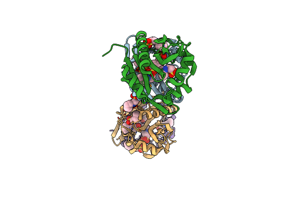
Tcar-Ssdna Complex Crystal Structure Reveals The Novel Ssdna Binding Mechanism Of The Marr Family Proteins
Organism: Staphylococcus epidermidis
Method: X-RAY DIFFRACTION Resolution:3.60 Å Release Date: 2014-03-19 Classification: TRANSCRIPTION/DNA Ligands: EDO, TRS |
 |
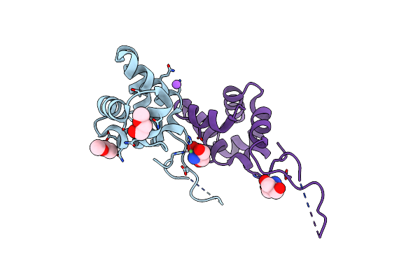
Structure Of Dna-Binding Domain Of The Response Regulator Saer From Staphylococcus Epidermidis
Organism: Staphylococcus epidermidis
Method: X-RAY DIFFRACTION Resolution:2.15 Å Release Date: 2014-01-29 Classification: TRANSCRIPTION |
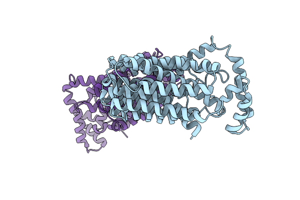 |
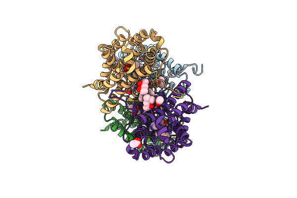
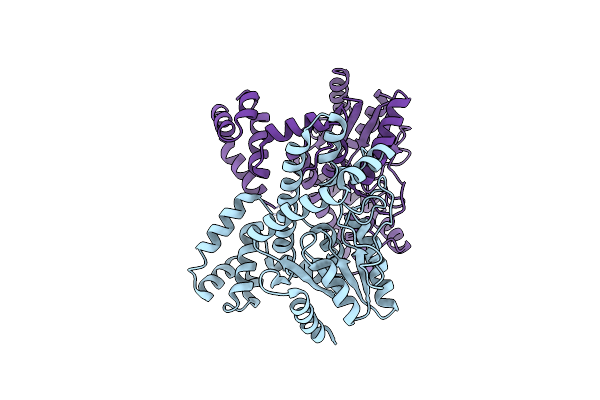
The Inward-Facing Structure Of The Glucose Transporter From Staphylococcus Epidermidis
Organism: Staphylococcus epidermidis (strain atcc 12228 / fda pci 1200)
Method: X-RAY DIFFRACTION Resolution:3.20 Å Release Date: 2013-10-16 Classification: transport protein, membrane protein |
 |
Organism: Staphylococcus epidermidis
Method: X-RAY DIFFRACTION Resolution:1.80 Å Release Date: 2013-09-04 Classification: HYDROLASE |
 |
The Crystal Structure Of A Possible An Iron-Binding (Periplasmic Solute-Binding) Protein From Staphylococcus Epidermidis Atcc 12228.
Organism: Staphylococcus epidermidis
Method: X-RAY DIFFRACTION Resolution:2.02 Å Release Date: 2013-08-28 Classification: Iron binding protein |
 |
Crystal Structure Of A Putative Thiaminase Ii (Se1693) From Staphylococcus Epidermidis Atcc 12228 At 1.65 A Resolution
Organism: Staphylococcus epidermidis
Method: X-RAY DIFFRACTION Resolution:1.65 Å Release Date: 2010-07-28 Classification: HYDROLASE Ligands: IMD, ACT, MPD, MRD, SO4 |
 |
The Crystal Structure Of The N-Terminal Domain Of A Rpir Transcriptional Regulator From Staphylococcus Epidermidis To 1.4A
Organism: Staphylococcus epidermidis
Method: X-RAY DIFFRACTION Resolution:1.40 Å Release Date: 2009-09-15 Classification: transcription regulator Ligands: CL, NI, NA, MXE, TRS |
 |
Crystal Structure Of A Probable Acetyltransferase From Staphylococcus Epidermidis Atcc 12228
Organism: Staphylococcus epidermidis atcc 12228
Method: X-RAY DIFFRACTION Resolution:2.70 Å Release Date: 2009-03-03 Classification: TRANSFERASE Ligands: NHE, FLC |
 |
Crystal Structure Of A Putative Nad(P)H:Fmn Oxidoreductase (Se1966) From Staphylococcus Epidermidis Atcc 12228 At 2.00 A Resolution
Organism: Staphylococcus epidermidis atcc 12228
Method: X-RAY DIFFRACTION Resolution:2.00 Å Release Date: 2009-03-03 Classification: OXIDOREDUCTASE Ligands: FMN, UNL, GOL, PGE, CA |
 |
The Crystal Structure Of A Functionally Unknown Conserved Protein From Staphylococcus Epidermidis Atcc 12228.
Organism: Staphylococcus epidermidis
Method: X-RAY DIFFRACTION Resolution:2.32 Å Release Date: 2009-01-27 Classification: structural genomics, unknown function |
 |
Organism: Staphylococcus epidermidis
Method: X-RAY DIFFRACTION Resolution:2.01 Å Release Date: 2009-01-13 Classification: structural genomics, unknown function |
 |
Crystal Structure Of Trna Delta(2)-Isopentenylpyrophosphate Transferase (Se0981) From Staphylococcus Epidermidis. Northeast Structural Genomics Consortium Target Ser100
Organism: Staphylococcus epidermidis atcc 12228
Method: X-RAY DIFFRACTION Resolution:2.70 Å Release Date: 2008-07-15 Classification: TRANSFERASE |