
Search Count: 141
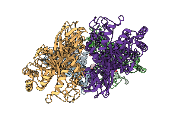 |
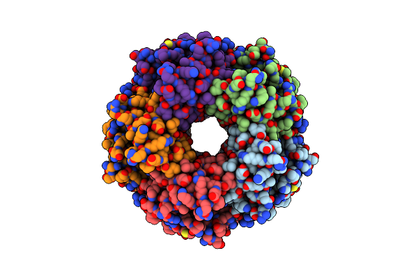
Glycoside Hydrolase Family 157 From Labilibaculum Antarcticum, Wild Type Semet Derivative (Lagh157)
Organism: Labilibaculum antarcticum
Method: X-RAY DIFFRACTION Resolution:2.44 Å Release Date: 2025-07-16 Classification: HYDROLASE Ligands: ACT, GOL, MLA |
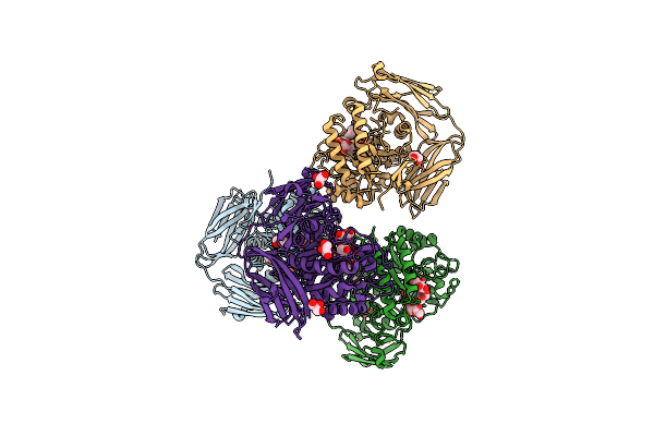 |
Glycoside Hydrolase Family 157 From Labilibaculum Antarcticum (Lagh157) E224A Mutant In Complex With Laminaritriose And Glucose
Organism: Labilibaculum antarcticum
Method: X-RAY DIFFRACTION Resolution:2.32 Å Release Date: 2025-05-21 Classification: HYDROLASE Ligands: EDO, BGC, GOL, PGE |
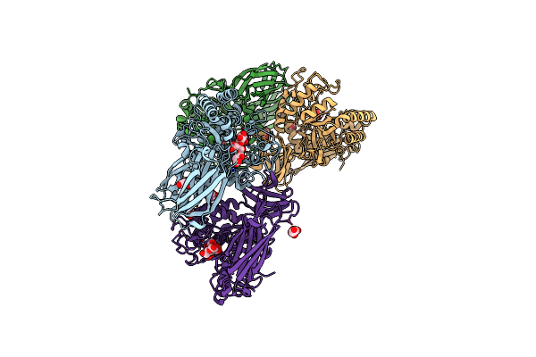 |
Glycoside Hydrolase Family 157 From Labilibaculum Antarcticum (Lagh157) In Complex With Laminaribiose
Organism: Labilibaculum antarcticum
Method: X-RAY DIFFRACTION Resolution:2.71 Å Release Date: 2025-05-21 Classification: HYDROLASE Ligands: MLA, GOL, BGC, PG4 |
 |
Cryo-Em Structure Of Coproheme Decarboxylase From Corynebacterium Diphtheriae In Complex With Heme B
Organism: Corynebacterium diphtheriae
Method: ELECTRON MICROSCOPY Release Date: 2025-02-05 Classification: OXIDOREDUCTASE Ligands: HEM |
 |
Cryo-Em Structure Of Apo Coproheme Decarboxylase From Corynebacterium Diphtheria.
Organism: Corynebacterium diphtheriae
Method: ELECTRON MICROSCOPY Resolution:2.27 Å Release Date: 2025-02-05 Classification: OXIDOREDUCTASE |
 |
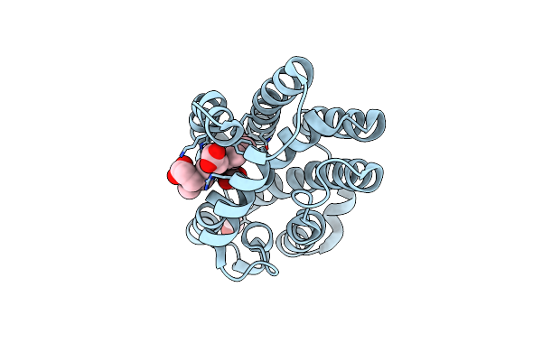
Crystal Structure Of Heme-Oxygenase From Corynebacterium Diphtheriae Complexed With Cobalt-Porphyrine (Humo-Co(Iii))
Organism: Corynebacterium diphtheriae
Method: X-RAY DIFFRACTION Resolution:1.15 Å Release Date: 2024-10-16 Classification: OXIDOREDUCTASE Ligands: COH, GOL, EDO, CIT, CL |
 |
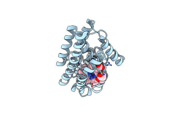
Crystal Structure Of Heme-Oxygenase From Corynebacterium Diphtheriae Complexed With Cobalt-Porphyrine (Humo-Co(Iii)) Flash-Cooled Under Co2 Pressure
Organism: Corynebacterium diphtheriae
Method: X-RAY DIFFRACTION Resolution:1.85 Å Release Date: 2024-10-16 Classification: OXIDOREDUCTASE Ligands: COH, EDO, CL, CO2 |
 |
Crystal Structure Of Heme-Oxygenase Mutant G139A From Corynebacterium Diphtheriae Complexed With Cobalt-Porphyrine (Humo-Co(Iii))
Organism: Corynebacterium diphtheriae
Method: X-RAY DIFFRACTION Resolution:1.40 Å Release Date: 2024-10-16 Classification: OXIDOREDUCTASE Ligands: COH, CL |
 |
Crystal Structure Of Heme-Oxygenase Mutant H20C From Corynebacterium Diphtheriae Complexed With Cobalt-Porphyrine (Humo-Co(Iii))
Organism: Corynebacterium diphtheriae
Method: X-RAY DIFFRACTION Resolution:2.30 Å Release Date: 2024-10-16 Classification: OXIDOREDUCTASE Ligands: COH |
 |
Crystal Structure Of Heme-Oxygenase Mutant I143K From Corynebacterium Diphtheriae Complexed With Cobalt-Porphyrine (Humo-Co(Iii))
Organism: Corynebacterium diphtheriae
Method: X-RAY DIFFRACTION Resolution:2.84 Å Release Date: 2024-10-16 Classification: OXIDOREDUCTASE Ligands: CO, COH, CL |
 |
Organism: Corynebacterium diphtheriae
Method: X-RAY DIFFRACTION Resolution:2.25 Å Release Date: 2023-07-05 Classification: TOXIN |
 |
Organism: Corynebacterium diphtheriae
Method: X-RAY DIFFRACTION Resolution:2.10 Å Release Date: 2023-07-05 Classification: TOXIN Ligands: APU |
 |
Organism: Corynebacterium diphtheriae
Method: X-RAY DIFFRACTION Resolution:2.00 Å Release Date: 2022-11-09 Classification: TOXIN Ligands: TRS |
 |
Organism: Corynebacterium diphtheriae
Method: X-RAY DIFFRACTION Resolution:2.00 Å Release Date: 2022-08-10 Classification: LIGASE Ligands: ANP, MYR, MG |
 |
Organism: Corynebacterium diphtheriae
Method: X-RAY DIFFRACTION Resolution:1.45 Å Release Date: 2022-04-20 Classification: OXIDOREDUCTASE Ligands: EDO, CL |
 |
Thiol-Disulfide Oxidoreductase Tsda, C129S Mutant, From Corynebacterium Diphtheriae
Organism: Corynebacterium diphtheriae
Method: X-RAY DIFFRACTION Resolution:1.10 Å Release Date: 2022-04-20 Classification: OXIDOREDUCTASE Ligands: PGE, EDO, CL |
 |
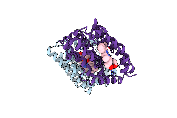
Crystal Structure Of Diphtheria Toxin Mutant Crm197 With A Disulphide Bond Replaced By A Cys-Acetone-Cys Bridge
Organism: Corynebacterium diphtheriae
Method: X-RAY DIFFRACTION Resolution:2.03 Å Release Date: 2022-04-06 Classification: TOXIN Ligands: 4Y8, EDO, PGE, ACT |
 |
Structure Of Coproheme Decarboxylase From Corynebacterium Dipththeriae W183Y Mutant In Complex With Coproheme
Organism: Corynebacterium diphtheriae
Method: X-RAY DIFFRACTION Resolution:2.15 Å Release Date: 2022-02-23 Classification: BIOSYNTHETIC PROTEIN Ligands: FEC, PGE, PEG |
 |
Structure Of Coproheme Decarboxylase From Corynebacterium Dipththeriae Y135A Mutant In Complex With Coproheme
Organism: Corynebacterium diphtheriae
Method: X-RAY DIFFRACTION Resolution:1.82 Å Release Date: 2022-02-23 Classification: BIOSYNTHETIC PROTEIN Ligands: FEC, PEG |
 |
Organism: Corynebacterium diphtheriae
Method: X-RAY DIFFRACTION Resolution:2.05 Å Release Date: 2020-11-18 Classification: TOXIN |