
Search Count: 87
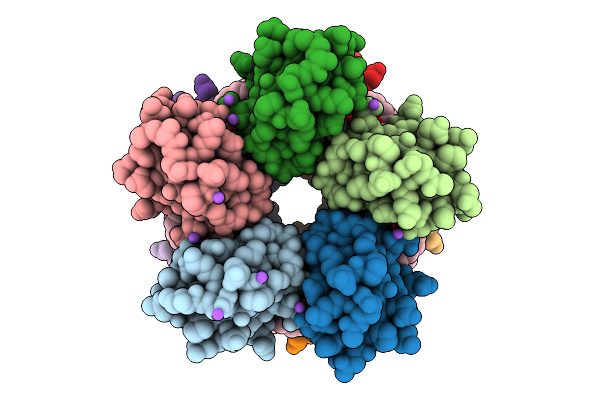 |
Organism: Desulfovibrio desulfuricans atcc 27774
Method: X-RAY DIFFRACTION Release Date: 2025-12-03 Classification: METAL BINDING PROTEIN Ligands: FE, A1CET, ACT, NA, 15P |
 |
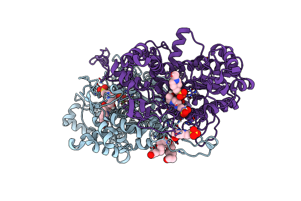
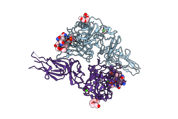
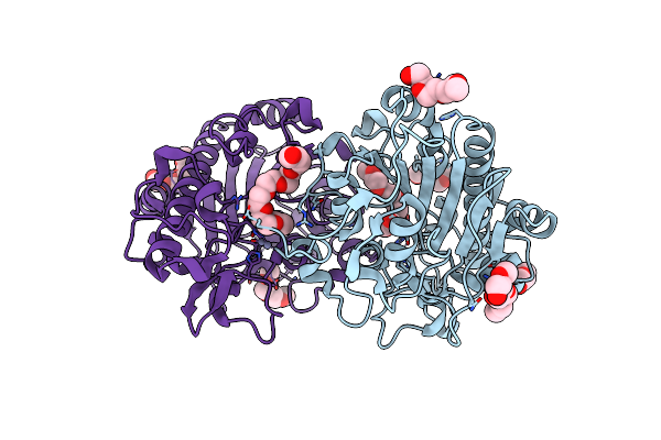
Crystal Structure Of Levansucrase From Pseudomonas Syringae In Complex With A Tetravalent Iminosugar
Organism: Pseudomonas syringae pv. actinidiae
Method: X-RAY DIFFRACTION Release Date: 2024-10-02 Classification: TRANSFERASE Ligands: MES, GOL, VXT, 15P, DMS |
 |
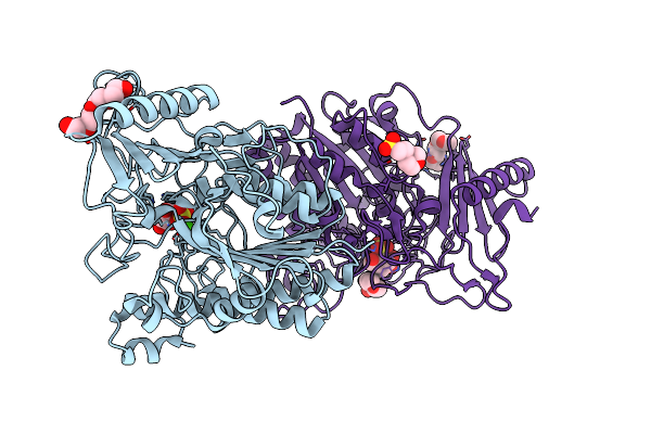
Apo X-Ray Crystal Structure Of Cyclophilin D With A Surface Entropy Reduction Mutation (K175I)
Organism: Homo sapiens
Method: X-RAY DIFFRACTION Resolution:1.87 Å Release Date: 2024-08-07 Classification: ISOMERASE Ligands: 15P, PEG, NA |
 |
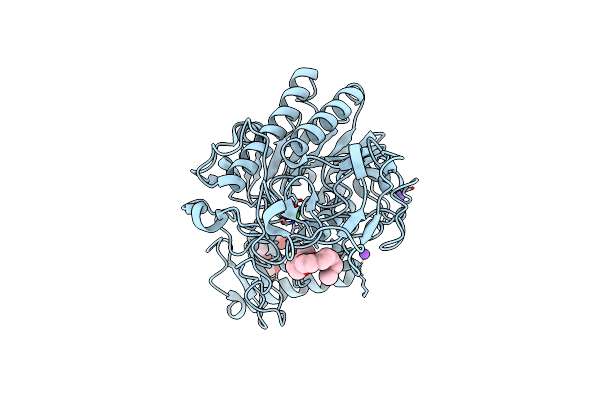
Organism: Vibrio sp. g15-21
Method: X-RAY DIFFRACTION Resolution:1.70 Å Release Date: 2022-11-02 Classification: HYDROLASE Ligands: ZN, MG, 15P, CL, EPE, PO4 |
 |
Sulfated Host Glycan Recognition By Carbohydrate Sulfatases Of The Human Gut Microbiota (Bt3796_S1_16)
Organism: Bacteroides thetaiotaomicron vpi-5482
Method: X-RAY DIFFRACTION Resolution:1.50 Å Release Date: 2022-06-29 Classification: HYDROLASE Ligands: P6G, G4S, CA, MES, 15P, CL |
 |
Sulfated Host Glycan Recognition By Carbohydrate Sulfatases Of The Human Gut Microbiota (Bt4631_S1_15)
Organism: Bacteroides thetaiotaomicron (strain atcc 29148 / dsm 2079 / nctc 10582 / e50 / vpi-5482)
Method: X-RAY DIFFRACTION Resolution:1.35 Å Release Date: 2022-06-29 Classification: HYDROLASE Ligands: 15P, P4K, CA, NA |
 |
Organism: Bacteroides thetaiotaomicron
Method: X-RAY DIFFRACTION Resolution:1.53 Å Release Date: 2022-03-02 Classification: UNKNOWN FUNCTION Ligands: CNC, CA, 15P, NA, MES |
 |
Structure Of Cytochrome C In Complex With A P-Benzyl-Sulfonato-Calix[8]Arene-Peg Pseudorotaxane
Organism: Saccharomyces cerevisiae s288c
Method: X-RAY DIFFRACTION Resolution:3.02 Å Release Date: 2021-10-20 Classification: OXIDOREDUCTASE Ligands: HEC, T8W, 15P |
 |
Exo-Beta-1,3-Glucanase From Moose Rumen Microbiome, Active Site Mutant E167Q/E295Q
Organism: Uncultured bacterium
Method: X-RAY DIFFRACTION Resolution:1.35 Å Release Date: 2021-01-13 Classification: HYDROLASE Ligands: 15P |
 |
Organism: Severe acute respiratory syndrome coronavirus 2
Method: X-RAY DIFFRACTION Resolution:1.95 Å Release Date: 2020-06-17 Classification: VIRAL PROTEIN Ligands: 15P |
 |
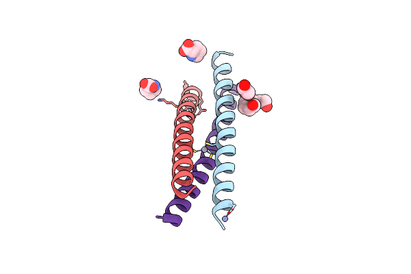
Crystal Structure Of A De Novo Three-Stranded Coiled Coil Peptide Containing A D-Leu In The Second Coordination Sphere Of A Non-Metalated Tris-Thiolate Binding Site
Organism: Synthetic construct
Method: X-RAY DIFFRACTION Resolution:1.40 Å Release Date: 2019-04-03 Classification: DE NOVO PROTEIN Ligands: ZN, 15P |
 |
Crystal Structure Of A Three-Stranded Coiled Coil Peptide Containing A Trigonal Planar Hg(Ii)S3 Site Modified By D-Leu In The Second Coordination Sphere
Organism: Synthetic construct
Method: X-RAY DIFFRACTION Resolution:1.84 Å Release Date: 2019-04-03 Classification: DE NOVO PROTEIN Ligands: ZN, PRO, 15P, CL, HG |
 |
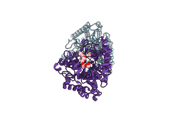
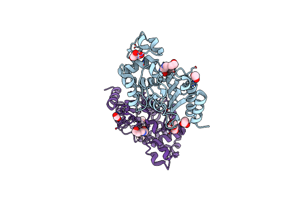
Structure Of S70A Blac From Mycobacterium Tuberculosis Obtained From Crystals Produced In The Absence Of Dtt
Organism: Mycobacterium tuberculosis
Method: X-RAY DIFFRACTION Resolution:1.19 Å Release Date: 2019-01-23 Classification: HYDROLASE Ligands: 15P, PEG, GOL |
 |
Structure Of Blac From Mycobacterium Tuberculosis Bound To The Trans-Enamine Adduct Derived From Clavulanic Acid.
Organism: Mycobacterium tuberculosis
Method: X-RAY DIFFRACTION Resolution:1.93 Å Release Date: 2019-01-23 Classification: HYDROLASE Ligands: ISS, ACT, GOL, 15P, CO2 |
 |
Structure Of Blac From Mycobacterium Tuberculosis Covalently Bound To Avibactam.
Organism: Mycobacterium tuberculosis
Method: X-RAY DIFFRACTION Resolution:1.62 Å Release Date: 2019-01-23 Classification: HYDROLASE Ligands: NXL, 15P |
 |
Organism: Homo sapiens
Method: X-RAY DIFFRACTION Resolution:2.00 Å Release Date: 2018-09-26 Classification: IMMUNE SYSTEM Ligands: GOL, 15P, MYR, PO4 |
 |
Organism: Drosophila melanogaster
Method: X-RAY DIFFRACTION Resolution:1.42 Å Release Date: 2018-06-20 Classification: SPLICING Ligands: 15P, SO4 |
 |
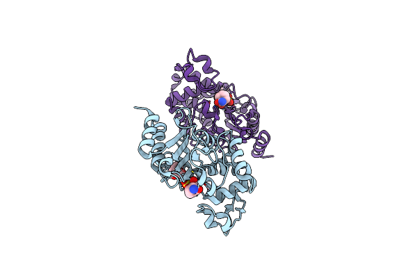
Crystal Structure Of The Intrinsic Colistin Resistance Enzyme Icr(Mc) From Moraxella Catarrhalis, Catalytic Domain, Thr315Ala Mutant Di-Zinc And Peg Complex
Organism: Moraxella sp. hmsc061h09
Method: X-RAY DIFFRACTION Resolution:1.50 Å Release Date: 2018-01-31 Classification: TRANSFERASE Ligands: ZN, 15P, CL |
 |
Crystal Structure Of The Intrinsic Colistin Resistance Enzyme Icr(Mc) From Moraxella Catarrhalis, Catalytic Domain, Thr315Ala Mutant Mono-Zinc And Phosphoethanolamine Complex
Organism: Moraxella sp. hmsc061h09
Method: X-RAY DIFFRACTION Resolution:1.66 Å Release Date: 2018-01-31 Classification: TRANSFERASE Ligands: ZN, OPE, SO4, 15P |
 |
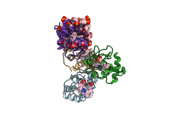
Crystal Structure Of The Broadly Neutralizing Influenza A Antibody Vrc 315 02-1F07 Fab.
Organism: Homo sapiens
Method: X-RAY DIFFRACTION Resolution:2.46 Å Release Date: 2017-12-13 Classification: IMMUNE SYSTEM Ligands: SO4, 15P, GOL |